训练用单针/双针带线【出售】-->外科训练模块总目录
0.5、1、2、3.5、5mm仿生血管仿生体 - 胸腹一体式腹腔镜模拟训练器
仿气腹/半球形腹腔镜模拟训练器
[单端多孔折叠]腹腔镜模拟训练器
「训练教具器械汇总」管理员微信/QQ12087382[问题反馈]
开启左侧

[资源] 腹腔镜治疗泌尿生殖道脱垂(图文演示)

[复制链接]
发表于 2016-7-21 10:31:21 | 显示全部楼层 |阅读模式

马上注册,结交更多好友,享用更多功能,让你轻松玩转社区。

您需要 登录 才可以下载或查看,没有账号?注册

×

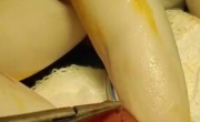
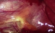
中文版:腹腔镜治疗泌尿生殖道脱垂(中文图文演示)

LAPAROSCOPIC   TREATMENT   OF   GENITOURINARY   PROLAPSE
Authors
JL Hoepffner, R Gaston
Abstract
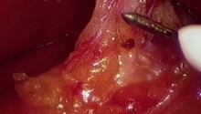
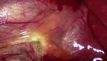
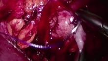
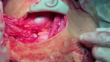
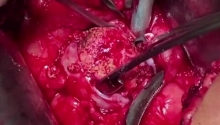
The description of the laparoscopic treatment of genitourinary prolapse covers all aspects of the surgical procedure used for the management of genital prolapse.
Operating room set up, position of patient and equipment, instruments used are thoroughly described. The technical key steps of the surgical procedure are presented in a step by step way: exposure, dissection, posterior prosthesis, closure, promontory, vesico-vaginal dissection, anterior mesh, promontory fixation, repair of peritoneum, drainage/closure.
Consequently, this operating technique is well standardized for the management of this condition.
 楼主| 发表于 2016-7-29 09:22:13 | 显示全部楼层
1. Introduction
Genitourinary prolapse is a frequent pathology in postmenopausal women. It is characterized by a deformation of the vagina caused by the prolapse of pelvic or abdominal organs. This prolapse results from weakness or damage to the natural aponeurotic or muscular support mechanisms.
 楼主| 发表于 2016-7-29 09:22:21 | 显示全部楼层
2. Anatomy
• Regional anatomy
Thorough knowledge of pelvic anatomy is essential for laparoscopic management of prolapse. This includes:
- position of the vagina in relation to the other pelvic organs;
- position of the promontory in relation to the iliac vascular pedicles and the ureter;
- location of the levator ani muscles.

[点击按纽展开更多->收起->回放]

1. Rectouterine (Douglas) pouch
After introducing the trocars into the peritoneal cavity, the parietal aspect of the peritoneum is incised:
- posteriorly, at the back of the rectouterine (Douglas) pouch to dissect the rectovaginal space, and laterally to expose the levator ani muscles;
- anteriorly, at the junction of the uterine isthmus and the anterior vaginal cul-de-sac, or opposite the fornix of the vagina in the case of a hysterectomy, to dissect the vesicovaginal space.

[点击按纽展开更多->收起->回放]

1. Anterior longitudinal ligament
2. Ureter
3. Median presacral vessels
The exposure of the anterior longitudinal ligament must follow the topography of the promontory with respect to the iliac vascular pedicles and the ureter. Care must be taken not to injure these organs during suturing of the prosthetic material.

[点击按纽展开更多->收起->回放]

1. Rectum
2. Puborectal muscle
3. Pubococcygeus muscle
4. Iliococcygeus muscle
Knowledge of the location of the levator ani muscles, the orientation of their fibers and their position with respect to the rectum, anal sphincter complex and vagina is essential.
 楼主| 发表于 2016-7-29 09:22:29 | 显示全部楼层
3. Indications
Indications
Laparoscopy can be performed to treat symptomatic genitourinary prolapse. The aim of the treatment is to control the clinical manifestations and to prevent complications.
Symptomatic prolapse:
- feeling of pelvic heaviness or fullness and low back pain;
- perception of a lump at the opening of the vulva;
- mucosal erosions.
Complications caused by prolapse:
- urinary tract infections;
- chronic bladder retention;
- bladder instability;
- dilatation of the upper urinary tract;
- renal insufficiency.

Contraindications
Anesthesia:
The contraindications are essentially those for general anesthesia.
Coagulation disorders:
Coagulation studies must be normal before operating.
Abdominal surgery:
It is possible to perform the procedure laparoscopically despite previous abdominal surgery. Laparoscopic adhesiolysis is mandatory in these cases.
Radiotherapy:
A history of radiotherapy in the pelvic region is not a contraindication, although it makes the dissection more difficult.
 楼主| 发表于 2016-7-29 09:22:35 | 显示全部楼层
4. Preop period
[点击按纽展开更多->收起->回放]

The diagnosis of genitourinary prolapse is purely clinical. There are several forms, depending on the organ involved. These different forms of prolapse are rarely isolated, but are combined in varying degrees.
1. Ureterocele:
On physical examination, the ureter and the bladder deform the anterior vaginal wall.
2. Cystocele:
On physical examination, the bladder deforms the anterior vaginal wall.
3. Rectocele:
On physical examination, the rectum deforms the posterior vaginal wall.
4. Hysterocele:
On physical examination, the uterus drops down, dragging along the vaginal vault.
5. Elytrocele:
On physical examination, the rectouterine pouch deforms the vaginal vault or the posterior vaginal wall.

[点击按纽展开更多->收起->回放]

Three grades of prolapse can be seen on physical examination. Grade 3 prolapse causes obvious physical discomfort. Grades 1 and 2 are generally asymptomatic. They are usually observed during physical examination in patients who consult for urinary incontinence.
Grade 1 is descent within the vagina.
Grade 2 is descent of the cervix to the introitus.
Grade 3 is descent of the uterus outside the introitus.

• Anesthetic work-up
The respiratory and cardiac conditions of the patient must permit general anesthesia. Coagulation studies must be normal and the urinalysis results negative before the procedure. The patient is informed of the operative risks and of the potential need for intraoperative conversion to an open procedure.

[点击按纽展开更多->收起->回放]

A resting
B straining
1. cystocele
2. rectocele
Urodynamic studies are often performed, especially in cases of coexisting incontinence.
MRI is used more commonly than colpocytogram for morphological exploration of the prolapse.
 楼主| 发表于 2016-7-29 09:22:42 | 显示全部楼层
5. Management
Management
Management of prolapse should comply with the following principles:
- repositioning of the organs while respecting their anatomical relationships;
- restoration or preservation of urinary and anal continence;
- maintenance of coital function;
- good long-term results.

Prolapse repair
Prolapse repair can be performed via 2 different approaches (Scali and Blondon, 1974):
- the high transabdominal approach;
- the low transvaginal approach.
Advocates of the transabdominal procedure have developed a laparoscopic approach (Cosson et al., 2000; Paraiso et al., 1999; Wattiez et al., 2001) to avoid a wide incision on the anterior abdominal wall.

Common objectives
The following objectives are shared by both conventional and laparoscopic abdominal procedures:
- interpose non-absorbable mesh between the anterior vaginal wall and the bladder, and between the posterior vaginal wall and the rectum. This material replaces the weakened means of natural support of the vagina, preventing recurrent prolapse.
- solidly attach this mesh to the anterior longitudinal ligament, at the level of the promontory. This tension-free attachment should reposition the vagina and restore its normal shape.

Specific objectives
The specific objectives of the laparoscopic approach are:
- attach the mesh in a minimal fashion to the posterior vaginal wall, to avoid the risk of vaginal necrosis;
- interpose graft material between the rectum and the vagina using a lateral attachment of the mesh on the levator ani muscles.
 楼主| 发表于 2016-7-29 09:22:49 | 显示全部楼层
6. Operating room
[点击按纽展开更多->收起->回放]

- general anesthesia;
- supine position;
- 30° Trendelenburg;
- perineum on the edge of the table to enable manipulation of the ribbon retractor;
- legs abducted to make the perineum accessible and to facilitate the positioning of the laparoscopic unit between the legs;
- prophylactic antibiotics are administered during anesthetic induction;
- urinary catheter.

[点击按纽展开更多->收起->回放]

1. The surgeon stands on the patient's left.
2. The assistant stands on the patient's right.
If needed, a second assistant manipulates the ribbon retractor and stands between the patient's legs or on the surgeon's left when acting as scrub nurse.
3. The scrub nurse stands on the surgeon's left with the instrument table to his or her left, or
between the patient's legs if acting as second assistant. If acting as first assistant, the scrub nurse
stands opposite the surgeon on the patient's right, with the instrument table to his or her right.
4. The anesthesiologist stands at the patient's head.

[点击按纽展开更多->收起->回放]

The operating table must be designed for a 30° Trendelenburg position.
Prolapse repair is often combined with surgical treatment for urinary incontinence. When a vaginal approach is required for this, the table must be equipped for a lithotomy position. If the patient’s legs can be easily attached to movable stirrups without contaminating the operative field, the initial draping can be maintained throughout the operation. Otherwise, the patient is repositioned after the prolapse repair, and re-draped.
 楼主| 发表于 2016-7-29 09:22:55 | 显示全部楼层
7. Trocar placement
[点击按纽展开更多->收起->回放]

Four trocars are usually necessary. The important landmarks are as follows:
- umbilicus;
- pubis;
- right anterior-superior iliac spine;
- left anterior-superior iliac spine.

[点击按纽展开更多->收起->回放]

The sizes and positions of the trocars are as follows:
A: 10 mm, infra-umbilical position
B: 5 mm, halfway between the umbilicus and the pubic symphysis
C: 5 mm, in the left iliac fossa, 2 to 3 cm medial to the anterior-superior iliac spine
D: 5 mm, in the right iliac fossa, 2 to 3 cm medial to the anterior-superior iliac spine
 楼主| 发表于 2016-7-29 09:23:09 | 显示全部楼层
8. Instruments
[点击按纽展开更多->收起->回放]

The instrumentation required for laparoscopic surgery is placed on the sterile instrument table to the right of the assistant or to the left of the scrub nurse, and includes the following:
1. two atraumatic graspers (one flat fenestrated and one narrow pair);
2. flat fenestrated bipolar forceps, which can also be used for grasping;
3. monopolar curved scissors;
4. laparoscopic needle holder;
5. suction-irrigation system;
6. urinary catheter;
7. specific ribbon retractor (flat, metal, rigid, 25 mm wide and 20 cm long, with one rounded endovaginal tip and one triangular tip);
8. non-absorbable mesh, either precut and preshaped, or cut to fit;
9. non-absorbable 0 braided suture, 26 mm needle, absorbable suture for the reconstruction of the peritoneum;
10. ultrasonic dissectors (optional).

[点击按纽展开更多->收起->回放]

The instruments are placed in the trocars as follows:
A: laparoscope
B: monopolar scissors or ultrasonic dissectors or needle holder
C: grasper or bipolar grasper
D: grasper or suction device
 楼主| 发表于 2016-7-29 09:23:16 | 显示全部楼层
9. Exposure
[点击按纽展开更多->收起->回放]

1. Uterus
2. Posterior vaginal wall
3. Rectouterine pouch
4. Rectum
The sigmoid colon is mobilized at the level of the intersigmoid recess to expose the rectouterine pouch. In rare cases, it is attached to the abdominal wall.
To expose the promontory, it is necessary to retract the sigmoid mesocolon towards the left.

[点击按纽展开更多->收起->回放]

The body of the uterus is suspended from the anterior abdominal wall with nylon 0 suture passed through the wall. This, along with the ribbon retractor, achieves tenting of the uterosacral ligaments and exposure of the anterior surface of the rectouterine pouch.

[点击按纽展开更多->收起->回放]

The Trendelenburg position maintains the intestinal loops outside the pelvic cavity.
您需要登录后才可以回帖 登录 | 注册

本版积分规则

丁香叶与你快乐分享

微信公众号

管理员微信

服务时间:8:30-21:30

站长微信/QQ

← 微信/微信群

← QQ

Copyright © 2013-2024 丁香叶 Powered by dxye.com  手机版 
快速回复 返回列表 返回顶部