19. Reference
Cosson M, Bogaert E, Narducci F, Querleu D, Crepin G. Promontofixation coelioscopique: resultats a
court terme et complications chez 83 patientes. J Gynecol Obstet Biol Reprod (Paris) 2000;29:746-
750.
Paraiso MF, Falcone T, Walters MD. Laparoscopic surgery for enterocele, vaginal apex prolapse and
rectocele. Int Urogynecol J Pelvic Floor Dysfunct 1999;10:223-9.
Scali P, Blondon J, Bethoux A, Gerard M. Les operations de soutenement-suspension par voie haute
dans le traitement des prolapsus vaginaux. J Gynecol Obstet Biol Reprod (Paris) 1974;3:365-78.
Wattiez A, Canis M, Mage G, Pouly JL, Bruhat MA. Promontofixation for the treatment of prolapse.
Urol Clin North Am 2001;28:151-7. |

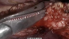
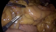
 腹腔镜下Whipple手术治疗胰头肿瘤
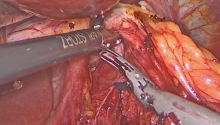
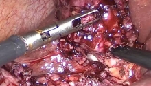
 腹腔镜治疗正中弓状韧带综合征(MALS)——
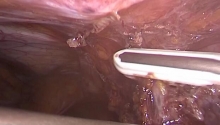
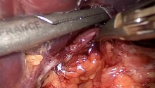
 腹腔镜治疗药物治疗失败后早期小肠梗阻复发
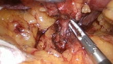
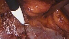
 腹腔镜全胃切除术联合吲哚菁绿(ICG)淋巴

 楼主
楼主


 显身卡
显身卡