训练用单针/双针带线【出售】-->外科训练模块总目录
0.5、1、2、3.5、5mm仿生血管仿生体 - 胸腹一体式腹腔镜模拟训练器
仿气腹/半球形腹腔镜模拟训练器
[单端多孔折叠]腹腔镜模拟训练器
「训练教具器械汇总」管理员微信/QQ12087382[问题反馈]
开启左侧

[资源] 肝右叶切除术(图文演示)

[复制链接]
 楼主| 发表于 2016-7-25 17:19:12 | 显示全部楼层
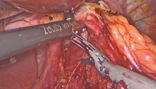
10. Approach/hepatic pedicle
[点击按纽展开更多->收起->回放]

Cholecystectomy is performed first in order to expose the superior portion of the right portal pedicle.
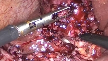
The triangle of Calot is exposed by retracting Hartmann’s pouch caudad and to the right. The anterior and posterior peritoneal leaves are divided down to the inferior surface of the liver. The cystic duct and the cystic artery are identified and separated. The cystic artery is divided first, after ligation or cauterization of the gallbladder neck. The cystic duct is subsequently divided.
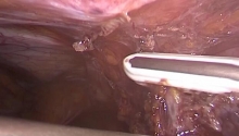
With the left hand, the first assistant pulls down the first duodenum to expose the anterior surface of the hepatoduodenal ligament. Segment 4 is lifted cephalad with a valve placed on its lower surface. This exposes the hilar plate, which is the reflection of the peritoneum of the porta hepatis on the capsule of segment 4, where the visceral peritoneum is slightly thickened.

• Hilar plate
[点击按纽展开更多->收起->回放]

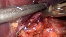
The peritoneum is opened at the level of the hilar plate parallel to the edge of segment 4, starting with the right part of the hilum. Only this right part should be divided.
It is best to use bipolar cauterization before dividing the peritoneum with sharp scissors.

[点击按纽展开更多->收起->回放]

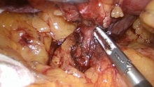
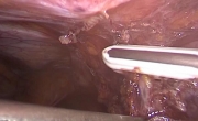
The decapsulated liver parenchyma appears at the upper part of the peritoneal incision. It is retracted slightly cephalad. The confluence of the right and left hepatic ducts is identified at the lower part of the incision, as well as the distal end of the right hepatic duct. This pulls the liver parenchyma off the right hepatic pedicle, which is lengthened artificially by this maneuver. The dissection of the right hepatic duct should be interrupted at this point.

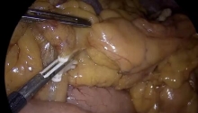
• Right branch of the portal vein
[点击按纽展开更多->收起->回放]

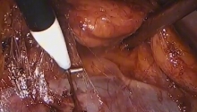
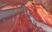
The peritoneum on the right inferior border of the porta hepatis is opened longitudinally, where the portal vein lies superficially. In order to facilitate the exposure, the first assistant slightly rotates the inferior part of the porta hepatis clockwise with the left hand. The portal vein lies just below the peritoneal layer.

[点击按纽展开更多->收起->回放]

The anterior and posterior surfaces of the portal vein are dissected free. The portal vein receives no tributaries in this segment.
The peritoneal incision is then extended cephalad along the right edge of the right portal vein. The superficial part of the hepatic pedicle is gently retracted cephalad with a small retractor. The dissection of the anterior surface of the portal vein and the exposure of the origin of the left portal vein is facilitated by a slight traction on the left portal vein. The areolar tissue situated between the main portal branches is cautiously retracted with the tip of blunt scissors to expose the origin of the right portal vein.

[点击按纽展开更多->收起->回放]

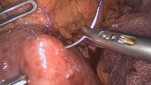
A tape can then be placed around the right portal vein. The dissecting instrument should be used with extreme caution around the right portal vein to avoid potential injury to the portal bifurcation or to the origin of the left portal vein.
If the right portal vein is not sufficiently freed, the portal bifurcation may be injured during the passage of the dissector, potentially resulting in massive bleeding.
The repair of this injury is very difficult. It is best to clamp the portal vein to decrease the blood flow.
A small branch supplying the right part of segment 1 originates from the right or posterior surface of the right portal branch near the portal bifurcation. This branch can be accidentally injured during isolation of the right portal vein. It is therefore preferable to ligate and divide it before taping.

[点击按纽展开更多->收起->回放]

The most common anatomical variations are either the division of the portal trunk into three branches or a segmental right portal vein arising from the left portal vein. If there are two different portal branches coursing towards the right liver, tape should be placed around them.

• Hepatic artery
[点击按纽展开更多->收起->回放]

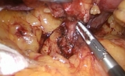
The right hepatic artery is searched for in the lymphatic tissue anterior to the right portal branch. This localisation is constant, even in cases of an anomalous pattern of the arterial tree (early division of the right hepatic artery, for example).

[点击按纽展开更多->收起->回放]

The lymphatic tissue is cautiously opened after performing bipolar cauterization. The right hepatic artery might already be divided into segmental or subsegmental branches. All of the remaining lymphatic tissue surrounding the artery is divided and the artery and its branches (if present) are placed on tape.
 楼主| 发表于 2016-7-25 17:19:21 | 显示全部楼层
11. Division/portal vessels I
[点击按纽展开更多->收起->回放]

Clamping of the right portal pedicle (right portal vein and right hepatic artery) results almost instantaneously in discoloration of the right liver and demarcation of the right and left lobes of the liver at the level of the interlobar fissure.

• Divisions
[点击按纽展开更多->收起->回放]

The right hepatic artery or its segmental or subsegmental branches are ligated and divided. This allows the surgeon to better expose the right portal vein and pursue its dissection up to the liver. It is then possible to identify and dissect the origin of the anterior and posterior branches of the right portal vein. This allows better visualization of this vessel, which can then be ligated more safely. Any residual lymphatic tissue anterior to the right branch of the portal vein is divided.

[点击按纽展开更多->收起->回放]

This operative step is crucial. Ligation of the right portal vein too close to its origin may result in a stenosis, leading to possible intraoperative or postoperative thrombosis of the portal vein with a high risk of hepatic failure and postoperative death. A light vascular clamp is placed on the origin of the right portal branch and one or two hemostatic clamps are placed on the distal part of the vein or at the origin of its segmental branches. The right portal vein is divided, taking care to leave a cuff on the proximal edge, which is oversewn with fine vascular suture. The distal part is ligated over the hemostatic clamp.

[点击按纽展开更多->收起->回放]

After the right portal branch has been divided, the right liver lobe remains attached to the hepatic pedicle by only the right hepatic duct and a band of fibrous tissue situated on the same plane as the duct extending towards the posterior edge of the liver (also called the posterior hilar plate). This band of fibrous tissue is divided with scissors. It often contains a small portal branch and more rarely a small bile duct.

[点击按纽展开更多->收起->回放]

Once the lower aspect of the right hepatic duct has been freed, it is possible to isolate it. The segmental right ducts join distally and thus the right hepatic duct is very short. Dissection should take place near the right and left hepatic duct confluence, because dissection of the right duct at a more proximal level is made difficult by the presence of segmental and subsegmental ducts.
A tape is placed around the right hepatic duct. The right hepatic duct is clamped distally at the level of the right and left hepatic duct confluence, avoiding any obstruction of the left hepatic duct, and proximally at the level of the segmental duct confluence. The right hepatic duct is divided and the distal and proximal ends suture- ligated with absorbable suture. This ligature is essential as any loosening of the distal stump may cause massive bile leakage. At this point, the right liver is completely detached from the hepatic pedicle.
 楼主| 发表于 2016-7-25 17:19:28 | 显示全部楼层
12. Division/portal vessels II
• Variation 1
▶
The right hepatic pedicle is transected “en masse” in the hilum.
Once the upper border of the right hepatic duct has been dissected, the inferior border of the right portal vein is exposed. An atraumatic clamp is passed between the hepatic parenchyma and the right hepatic duct, around the portal vein. The right hepatic pedicle is isolated with a tape. This maneuver can be dangerous, as bleeding may occur and distal vascular control is impossible to obtain in the deeper part of the operative field. After slight anterior traction on the tape, a mechanical stapler is applied to the entire right hepatic pedicle. Several firings of the mechanical stapler may be necessary to divide the right pedicle. It has been suggested that blood loss is less after “en masse” ligation of the right hepatic pedicle than after an isolated division of these vessels, ducts and bile duct.

• Variation 2
▶
Intraparenchymal division of the right hepatic pedicle is performed.
The principle of this technique is to divide the vessels and ducts as far from the porta hepatis as possible, thereby reducing the risk of injury to a vessel or main duct linked to the remaining liver, particularly in case of anatomical variations. The right hepatic artery and the right portal vein are dissected and isolated as described previously. The right hepatic duct is only partially dissected. The vessels are clamped. Parenchymal transection is carried out until the biliary confluence is reached. Transection should be performed slightly to the right of the usual plane. The vessels and ducts are dissected within the parenchyma and divided separately. This approach is not considered to be safer than extraparenchymal division. In the latter technique, division is performed on segmental branches that are sometimes difficult to isolate. In addition, the division of the pedicle within the parenchyma pushes the line of transection slightly to the right, making the last parenchymal division and right hepatic vein approach more difficult.

• Variation 3
Quite frequently, a subsegmental or segmental duct drains separately into the common hepatic duct slightly below the confluence of the right and left hepatic ducts. This variation should not be taken into account in the technique. The distance separating the right hepatic duct and this other branch is short enough for the clamp to be placed on the two ducts without the need to identify them.
 楼主| 发表于 2016-7-25 17:19:35 | 显示全部楼层
13. Dissection/suprahepatic vein
[点击按纽展开更多->收起->回放]

The cellular tissue between the right and middle hepatic vein is gently retracted with the tip of blunt scissors and divided. The left border of the right hepatic vein is exposed before it enters the IVC. The right liver is rotated towards the left upper quadrant in order to expose the IVC.

• Dissections
[点击按纽展开更多->收起->回放]

The IVC is situated above the adrenal gland posterior to the liver. Its right surface is covered with a fibrous ligament called the hepatocaval ligament, which extends between the posterior border of the liver and the posterior surface of the vena cava. While it is very thin in most patients, it is thick in more than 20% of patients.
This ligament is divided to expose the right surface of the superior part of the IVC. It is sometimes easy to separate it from the vein, but in about 15% of cases, it closely adheres to the venous wall, making the procedure difficult to perform.

[点击按纽展开更多->收起->回放]

The right border and the right half of the anterior surface of the IVC are dissected. A certain number of small hepatic branches (also called Spiegelian veins) are observed from the posterior surface of the liver to the IVC; they are ligated and divided.
This dissection should be performed gently since tearing of a branch may cause massive bleeding that is difficult to control before complete right mobilisation of the liver from the IVC. If hemostasis is necessary, it should be achieved with an X-shaped stitch using fine suture.

[点击按纽展开更多->收起->回放]

Once the hepatocaval ligament has been divided, the inferior border of the right hepatic vein is exposed. The left border of the vein is isolated using gentle caudad to cephalad dissection. An atraumatic dissector is passed around the vein caudad to cephalad so a tape can be placed. This maneuver must be performed with the utmost care to avoid injuring the middle or right hepatic veins. If complete isolation of the right hepatic vein is made impossible by an obstacle, further caudad to cephalad dissection of the left border of the vein should be performed. In our experience, it was possible to completely isolate the right hepatic vein with a tape in 99.5% of right liver resections, even in cases of voluminous tumors.

• Dangers
[点击按纽展开更多->收起->回放]

Injury to a main hepatic vein may cause major bleeding and gas embolism.
The injury is packed with sponges to temporarily stop bleeding and stabilize the state of the patient. Hemostasis can later be performed with one or several interrupted stitches with fine suture. The pressure is low in the hepatic veins and as a result bleeding can be easily controlled.
Note: Hemostasis performed too rapidly when the exposure is poor can aggravate bleeding and venous injuries.

[点击按纽展开更多->收起->回放]

Dissection of the hepatocaval ligament from the right hepatic vein involves a risk of injury to the hepatic vein or the IVC.
In about 5% of patients, this ligament is replaced by a bridge of hepatic parenchyma that should be divided. This bridge usually contains a small portal branch and a bile duct. This parenchymal bridge may be up to 10 mm thick.

[点击按纽展开更多->收起->回放]

In about 20% of patients, there is another large right hepatic vein close to the inferior edge of the liver. This is the right inferior hepatic vein. It is easier to divide this inferior hepatic vein between two hemostatic clamps to complete the dissection of the IVC.
 楼主| 发表于 2016-7-25 17:19:45 | 显示全部楼层
14. Transection/parenchyma
[点击按纽展开更多->收起->回放]

Before dividing the liver parenchyma, the fibrous capsule of the liver is incised over its entire length using electrocautery, along the interlobar fissure that can be distinguished by ischemic demarcation caused by vessel division. It is preferable to remain 2 mm to the right of this line so the parenchymal division can be done on the right flank of the middle hepatic vein, which is preserved, and to remain on the transection surface. The incision of the capsule starts from the superior surface of the liver on the left edge of the right hepatic vein over the hilum, and skirts around the anterior border of segment 1 (at its narrowed part). It is then continued cephalad to the posterior surface of the liver, along the IVC, up to the left edge of the right hepatic vein.

• Operative technique
[点击按纽展开更多->收起->回放]

The parenchymal division is started at the anterior border of the liver. It follows the interlobar fissure while keeping slightly to the right of the middle hepatic vein. The division is performed step-by-step through the entire thickness of the liver, which opens out like a book. Once the hilus is reached, division should start again from the anterior border of segment 1 and be pursued cephalad towards the superior and posterior surface of the liver.

[点击按纽展开更多->收起->回放]

On the superior part of the parenchymal division, the left border of the right hepatic vein is approached. The peritoneal leaf covering it is opened and the vein is skeletonized. A vascular clamp is placed on the vein at its precise junction with the IVC. The right hepatic vein is then divided.

[点击按纽展开更多->收起->回放]

After removing the operative specimen, the right hepatic vein stump is oversewn with fine vascular suture. Alternatively, the vein can be closed with a vascular stapler. Special care must be taken to achieve perfect hemostasis in closing the lateral wall of the IVC.
 楼主| 发表于 2016-7-25 17:19:57 | 显示全部楼层
15. Transection techniques
[点击按纽展开更多->收起->回放]

Numerous vessels and bile ducts run through the liver parenchyma. In order to transect the parenchyma without causing bleeding or bile leakage, it should be divided while preserving the blood vessels and ducts. These are ligated and divided as they are exposed.
Finger fracture or digitoclasty (crushing the hepatic parenchyma between the thumb and the forefinger) was the first procedure used. This technique is rather traumatic and exposes smaller vessels to injury.
Finger fracture has been replaced by vascular clamp-assisted parenchymal dissection, also referred to as kellyclasty. This is a simple method. It is essential to progress slowly and to crush only thin parenchymal fragments, to avoid bleeding and injuries to small vessels.

[点击按纽展开更多->收起->回放]

Ultrasound emission from the tip of a handpiece gently dissociates the liver tissue.
Ultrasonic dissectors are provided with an irrigation-suction device that is used to continually cleanse the dissection plane. The power of ultrasound must be adjusted to the firmness of the parenchyma. When the adjustment is correctly done, the risk of vascular or biliary injury is very low. The few properly designed studies comparing kellyclasty to ultrasonic dissection suggest that there is less blood loss with ultrasonic dissectors than with kellyclasty.

[点击按纽展开更多->收起->回放]

Dissociation of liver tissue may also be performed with a high pressure water jet (2 to 3 bars). The saline solution, blood and micro tissue debris are aspirated through a suction cannula incorporated into the handpiece. The line of transection is flat and smooth. The pressure of the jet should be adapted to the firmness of the parenchyma. When the adjustments are properly done, even small vascular and biliary structures are preserved.

• Coagulation-division
[点击按纽展开更多->收起->回放]

These techniques permit “en masse” coagulation of a liver tissue fragment and the small pedicles running through it until necrosis and spontaneous division occur.
The techniques that are currently available to surgeons use the heat produced by very high frequency vibrations applied to the blades of scissors. Only vessels and bile ducts measuring less than 3 mm in diameter may be divided using this technique.

[点击按纽展开更多->收起->回放]

Division of intraparenchymal pedicles can be performed using fine suture, metal or absorbable clips, or bipolar coagulation. Bipolar coagulation is used more often nowadays for most pedicles. In a right liver resection, only a small number of collaterals of the middle hepatic vein need to be ligated or clipped.

[点击按纽展开更多->收起->回放]

If bleeding occurs during this division, it is most often due to the tearing or avulsion of a collateral from the middle hepatic vein.
It is important to avoid poorly controlled reflexes such as abruptly separating the two parts of the liver or thrusting the suction-irrigation device into the dissection plane to better expose the hemorrhagic zone. These two maneuvers aggravate injuries and bleeding.
In the absence of a clear understanding of bleeding mechanisms, it is often better to place sponges between the two parts of the liver and exert manual pressure for one to two minutes. This can stop bleeding, or at least limit it, to permit better exposure and hemostasis.
 楼主| 发表于 2016-7-25 17:20:05 | 显示全部楼层
16. Control/transection
• Simple transection
[点击按纽展开更多->收起->回放]

When the hepatic transection is uneventful and the division of biliary and vascular pedicles is done step by step, the cut surface of the liver is dry at the end of the resection and requires no additional maneuver. A thorough examination is still necessary. Hemostasis and biliostasis are checked one last time with the remaining liver back in place, as exposure maneuvers may conceal bleeding or minimal bile leaks.

• Bleeding
▶
Superficial bleeding is stopped by bipolar cautery. Deeper bleeding, whether arterial or portal, is stopped by suture ligatures with fine suture. The tightening of these stitches should be gradual so as not to tear the hepatic parenchyma.
Hepatic venous bleeding usually stops after simple compression.

• Bile leak
When a bile leak is detected near the left hepatic duct, its origin should be absolutely identified and the wound repaired by fine X-shaped monofilament absorbable sutures.

• Complicated transection
• Risks
When the parenchymal division is long and difficult, especially in diseased livers with steatosis or fibrosis, or in patients who have undergone chemotherapy, the dissection plane can be hemorrhagic at the end of the resection.

• Limited bleeding
▶
In the absence of major bleeding, either arterial or portal, it is best to apply compression with sponges and moderate manual pressure for a few minutes. Complete hemostasis can usually be achieved. Placing sponges soaked with coagulation factors on the area can help obtain permanent hemostasis.

• Bile leaks
The injection of a dye in the bile ducts via a cystic cannula does not improve the screening for a bile leak in the region and is therefore not recommended. An intraoperative cholangiography performed via the cystic duct is useful only when major injury to the upper biliary confluence is suspected. This is very unlikely during a right hepatectomy if the technique has been carefully followed.

• Abdominal drainage
Abdominal drainage after hepatic resection has evolved in the same manner as it has for other types of abdominal surgery. If the surgical procedure is performed properly, the ligature of the main vessels is reliable and the cut surface of the liver is dry, abdominal drainage is not useful. Many surgeons still prefer to leave an abdominal drain. A small aspiration drain is most convenient.

• Variation
The use of biological glue sprayed onto the line of transection at the end of the procedure is controversial. In our experience, it does not allow the surgeon to seal a bile leak or to stop bleeding that compression alone cannot manage.
 楼主| 发表于 2016-7-25 17:20:13 | 显示全部楼层
17. Postop period
The IV is left in place until bowel function is restored. This generally occurs 2 to 4 days after the procedure. Oral food intake can be started as soon as the bowel function resumes. Although antibiotic prophylaxis is generally advised, there is no evidence that it is useful. It is preferable to wait for 24 hours after surgery before starting anticoagulant therapy, to avoid bleeding at the parenchymal dissection plane. Early ambulation of the patient is recommended, as for all surgical procedures. Postoperative follow-up is essentially clinical.
 楼主| 发表于 2016-7-25 17:20:21 | 显示全部楼层
18. Results
Liver resection is a well-tolerated procedure in patients with a normal liver, as long as the volume of remaining parenchyma is greater than 30% of whole liver volume.

Liver function tests
Postoperative alteration of liver function test results is early and short-term. After major liver resections such as right or extended right hepatectomy, serum bilirubin concentration can rise to 50 micromoles/L and is at its highest level on the second or third postoperative day. Prothrombin time decreases to a mean of 40%. SGOT and SGPT levels increase to about 5 to 10 times their normal value. Increase in liver transaminases depends not only on the size of the parenchymal resection but also on intraoperative bleeding and the duration of portal triad clamping. Changes in serum bilirubin concentration, prothrombin time and transaminases beyond these values or for a more prolonged period of time are predictive of postoperative complications and should prompt investigations.

Operative mortality
Operative mortality now averages 1% in patients with a normal liver parenchyma. It is higher following major liver resection and resection of malignant tumors than following minor resection and treatment of benign tumors (Belghiti et al., 2000). Estimated mortality in living donor liver transplantation, the best case for liver resection, is less than 0.2% (Renz and Roberts, 2000).

Other complications
The overall complication rate still averages 40%, and includes pneumonia, urinary tract infection and peripheral venous thrombosis. Severe complications occur in about 15% of patients. Abdominal complications occur in about 10% of cases. They require reoperation in about 3% of patients who undergo liver resection. The most frequent complications are bleeding from the remaining liver or from small vessels, bile leakage and subphrenic abscess. Right pleural effusion is particularly common and may occur independently of a subphrenic abscess or hematoma.
 楼主| 发表于 2016-7-25 17:20:43 | 显示全部楼层
19. Reference
Belghiti J, Hiramatsu K, Benoist S, Massault P, Sauvanet A, Farges O. Seven hundred forty-seven hepatectomies in the 1990s: an update to evaluate the actual risk of liver resection. J Am Coll Surg 2000;191:38-46.
Renz JF, Roberts JP. Long-term complications of living donor liver transplantation. Liver Transpl
2000;6:S73-6.
您需要登录后才可以回帖 登录 | 注册

本版积分规则

丁香叶与你快乐分享

微信公众号

管理员微信

服务时间:8:30-21:30

站长微信/QQ

← 微信/微信群

← QQ

Copyright © 2013-2024 丁香叶 Powered by dxye.com  手机版 
快速回复 返回列表 返回顶部