训练用单针/双针带线【出售】-->外科训练模块总目录
0.5、1、2、3.5、5mm仿生血管仿生体 - 胸腹一体式腹腔镜模拟训练器
仿气腹/半球形腹腔镜模拟训练器
[单端多孔折叠]腹腔镜模拟训练器
「训练教具器械汇总」管理员微信/QQ12087382[问题反馈]
开启左侧

[资源] 腹腔镜Nissen胃底折叠术治疗胃食管反流病(图文演示)

[复制链接]
 楼主| 发表于 2016-7-24 07:34:49 | 显示全部楼层
10. Dissection/cardioesophageal junction
• Principles
The intervention begins with the mobilization of the gastroesophageal junction.
The successive dissection steps allow to identify the essential anatomical structures: the diaphragmatic crura, the esophagus, the vagus nerves, the mediastinal pleura and the aorta.

[点击按纽展开更多->收起->回放]

The pars condensa (superior part) of the lesser omentum is opened whilst making sure to preserve the hepatic branch of the vagus nerve and the left hepatic artery. This phase helps to identify the right diaphragmatic crus, the essential anatomic landmark that needs to be identified before beginning the dissection of the esophagus.

[点击按纽展开更多->收起->回放]

Incision of the phrenoesophageal membrane is continued on the medial relief of the right crus.
Non-traumatic dissection of the cleavage plane that is found internal to the right crus allows to identify the position of the esophagus. Finding that area is facilitated by lifting the distal portion of the esophagus and the cardia anteriorly and to the left. That step allows access to the crura, the posterior edge of the esophagus and the posterior vagus nerve.

[点击按纽展开更多->收起->回放]

Incision of the phrenoesophageal membrane and dissection of the left crus:
Once the position of the esophagus is identified, the phrenoesophageal membrane is opened transversally along the anterior border of the hiatal orifice and towards the left crus. At that level, a blunt dissection of the internal aspect of the crus is recommended to avoid damage to the anterior vagus nerve that may be found proximal to it.
The dissection is then continued towards the most posterior part of the crus to prepare the retroesophageal dissection.

• Dissection/posterior aspect of esophagus
[点击按纽展开更多->收起->回放]

Dissection of the posterior aspect of the esophagus allows to create the retroesophageal window. It is carried out from right to left posterior to the esophagus and in contact with the external border of the left crus.
The posterior vagus nerve is identified and retracted against the wall of the esophagus.
The window is created when the fat pad of the gastrosplenic ligament and/or the upper aspect of the spleen are visible.
Identification of such landmarks is essential during this dissection: the right crus and the left crus help to guide the dissection. A dissection performed above the left crus could lead to a pleural injury.
The adjacent anatomical structures must be clearly identified.

[点击按纽展开更多->收起->回放]

A Penrose drain or a loop, held with a grasper by the assistant (through trocar D), is passed through the enlarged retroesophageal window, and then placed around the esophagus. It permits to avoid any traumatic grasping of the organs and helps to mobilize the gastroesophageal junction in different directions during the next surgical steps.
 楼主| 发表于 2016-7-24 07:34:57 | 显示全部楼层
11. Mobilization of esophagus
• Objectives
- to free the lower esophagus and cardia on their anterior and posterior aspects;
- to create a retroesophageal window of sufficient size to allow passage of the fundus;
- to sufficiently mobilize the esophagus in the abdominal cavity (and obtain a segment of esophagus more than 2 cm long inside the abdominal cavity in a tension-free manner).

[点击按纽展开更多->收起->回放]

Dissection of the esophagus into the mediastinum lengthens the abdominal portion of the esophagus. This portion should be at least 2 cm long without tension.
This dissection can be extended up to the pulmonary veins, if not to a greater length in some circumstances. The vagus nerves must systematically be identified and protected.
If the cardia cannot be lowered despite this dissection, further dissection of the esophagus into the mediastinum should be performed. A Collis gastroplasty procedure (i.e. elongation/lengthening of the esophagus) is advisable in the case of a short esophagus.

[点击按纽展开更多->收起->回放]

Division of the posterior attachments of the cardia on the diaphragm allows to enlarge the retroesophageal window. The possibility of a left diaphragmatic artery crossing through that space must be kept in mind.
 楼主| 发表于 2016-7-24 07:35:05 | 显示全部楼层
12. Mobilization of gastric fundus
[点击按纽展开更多->收起->回放]

The surgical technique described includes mobilization of the gastric fundus, which is used to create the antireflux valve.
Division of the gastrosplenic ligament, of two or three short gastric vessels, and in certain cases, of the posterior fundic artery makes up the essentials of this operative time.

[点击按纽展开更多->收起->回放]

The gastrosplenic ligament is exposed using traction on the fundus of the stomach towards the patient’s right (trocar B) and lateral traction on the gastrosplenic ligament towards the patient’s left (trocar D).
The ligament is first divided to its cephalad origin where some fat folds may be found. Monopolar or bipolar cautery, ultrasonic scissors or a vessel sealing device may be used through trocar E.

[点击按纽展开更多->收起->回放]

Incision of the gastrosplenic ligament gives access to the omental sac.
Cephalad traction on the fundus (trocar B) exerts tension on the short gastric vessels and posterior attachments of the stomach. Retraction of the body of the stomach caudally and to the patient’s right helps to expose these structures (trocar D).
Two to three short gastric vessels are divided. Division of the posterior layer of the gastrosplenic ligament is continued cephalad. It is then followed by division of the gastrophrenic ligament. At the end of dissection, the left crus is entirely visible.
 楼主| 发表于 2016-7-24 07:35:12 | 显示全部楼层

[点击按纽展开更多->收起->回放]

Plasty of the diaphragmatic opening should be performed routinely as it not only contributes to restoring one of the elements of the antireflux barrier but also stabilizes the antireflux wrap into the abdominal cavity.
The esophagus is retracted distally from the crura using the loop that is passed around the gastroesophageal junction (trocar D). Grasper and needle holder are used through trocars B and E.
Interrupted non-absorbable sutures are placed on the crura to repair the hiatal defect posterior to the esophagus.
At the beginning of the experience, it is recommended to place a 55 French bougie into the esophagus to accurately size the esophagus and avoid any strangulation.
Sutures may be placed anterior to the esophagus in cases of very large hiatal defects.
If the crura are fragile, Teflon pledgets might be useful to apply sutures.
Prosthetic reinforcement may be used when needed.
 楼主| 发表于 2016-7-24 07:35:19 | 显示全部楼层
14. Creation of the fundoplication
[点击按纽展开更多->收起->回放]

For Nissen fundoplications, the anterior and posterior walls of the gastric fundus are mobilized contrarily to Nissen-Rossetti fundoplications where the anterior wall only is mobilized.

[点击按纽展开更多->收起->回放]

The esophagus is pulled up using the loop.
The posterior wall of the gastric fundus is brought to the right of the esophagus through the posterior esophageal window. The anterior fundic wall slides towards the posterior aspect of the esophagus.
Once tailored, the valve should be sufficiently stable without tension towards the spleen. When tension is still encountered, a wider mobilization of the stomach is to be performed to avoid any tension on the valve and prevent any twisting of the lower esophagus postoperatively.

[点击按纽展开更多->收起->回放]

The valve should be positioned at the level of the Z line, 1 to 1.5 cm above the anatomical junction. It must always be positioned way above the omental folds.
The valve should be tailored using the gastric fundus. Caution must be taken not to fashion the valve with the body of the stomach as there is a risk of partitioning the stomach into two parts.
In case of doubt about the positioning of the fundoplication (especially in the case of short esophagus), it is recommended to perform an intraoperative esophagoscopy.

[点击按纽展开更多->收起->回放]

The two parts of the valve are joined together using three non-absorbable sutures.
The first most cephalad suture does not attach the valve to the esophagus. It helps to check the fundoplication, the absence of plications of gastric walls, and controls correct positioning of the valve on the esophagus.
The next two sutures complete the fundoplication and fix it to the anterior aspect of the esophagus and to the right of the anterior vagus nerve. The 360-degree Nissen fundoplication is 1.5 to 2 cm in length and height. It is maintained in position by three interrupted, non-absorbable, U-shaped sutures (0.0). An additional stitch may be used to fix the left part of the valve to the left border of the esophagus at the insertion of the phrenoesophageal membrane.
 楼主| 发表于 2016-7-24 07:35:25 | 显示全部楼层
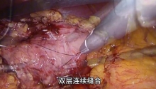
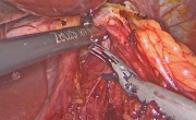
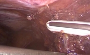
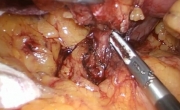
15. Intraoperative complications
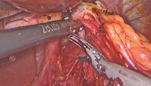
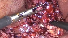
Perforations
Frequency:
- rare (approximately 1%),
- dangerous if not immediately recognized intraoperatively as they carry a mortality of 20% to 50%15,17.
Mechanisms:
- placement of a bougie or nasogastric tube;
- traumatic manipulations of the esophagus sometimes attenuated by an inflammation;
- blind dissection in the absence of fixed anatomic landmarks.
What to do:
- primary closure of the perforation covered with the fundoplication.
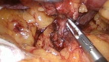
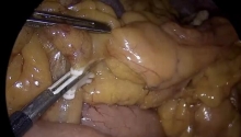
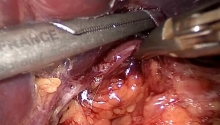
Hemorrhage
Frequency:
- rare, often mild, not requiring transfusions.
Mechanism:
Bleeding could originate from:
a. the abdominal wall, at a trocar insertion site;
b. a short gastric vessel;
c. a diaphragmatic artery, especially at the level of the left crus;
d. hepatic trauma with a retractor or instrument;
e. a splenic laceration.
What to do:
a. suture ligation,
b. and c. hemostatic control using bipolar coagulation,
d. compression with a retractor or use of argon beam coagulator,
e. use of argon beam coagulator or fibrin glue.
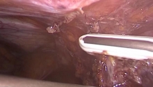
Pneumothorax
Frequency:
- CO2 pneumothorax is a specific but benign complication of the laparoscopic approach,
- its incidence is approximately 3%, but is likely to be underestimated.
Mechanism:
It is caused by rupture of the pleura, more often on the left than on the right one, during a prolonged mediastinal dissection.
What to do:
- the treatment of the pneumothorax involves modification of the ventilation parameters with the addition of PEEP (positive end expiratory pressure),
- thoracic drainage is not necessary: the postoperative chest radiograph is often normal as the CO2 is rapidly absorbed when insufflation is discontinued.

Emphysema
Frequency:
- rare
Mechanism:
- mediastinal and/or subcutaneous emphysema can present occasionally during or after an operation when the hiatal dissection is too deep or prolonged.
What to do:
- the first therapeutic measure is to adjust the ventilation rate with or without a reduction of the insufflated pressure.

Vagus nerve trauma
Frequency:
- rarely reported, as it is often unrecognized.
Mechanism:
- the nerve can inadvertently be divided using electrocautery or damaged by diffusion of electrocautery current:
A: during the dissection of the posterior aspect of the esophagus for the posterior vagus nerve.
B: during the dissection of the phrenoesophageal membrane for the anterior vagus nerve.
What to do:
- prevention: careful dissection and identification of the 2 nerves.
 楼主| 发表于 2016-7-24 07:35:33 | 显示全部楼层
16. Closure
The peritoneal cavity is rinsed with warm normal saline.
No drains are placed.
The trocars are removed and the skin openings are closed with staples.
Fluid intake begins on the same day.
Solid intake begins on the first postoperative day.
The patient is usually discharged on the second postoperative day.
 楼主| 发表于 2016-7-24 07:35:40 | 显示全部楼层
17. Bibliographic references
您需要登录后才可以回帖 登录 | 注册

本版积分规则

丁香叶与你快乐分享

微信公众号

管理员微信

服务时间:8:30-21:30

站长微信/QQ

← 微信/微信群

← QQ

Copyright © 2013-2024 丁香叶 Powered by dxye.com  手机版 
快速回复 返回列表 返回顶部