训练用单针/双针带线【出售】-->外科训练模块总目录
0.5、1、2、3.5、5mm仿生血管仿生体 - 胸腹一体式腹腔镜模拟训练器
仿气腹/半球形腹腔镜模拟训练器
[单端多孔折叠]腹腔镜模拟训练器
「训练教具器械汇总」管理员微信/QQ12087382[问题反馈]
开启左侧

[资源] 腹腔镜全直肠系膜切除术(TME)癌症(图文演示)

[复制链接]
 楼主| 发表于 2016-7-24 15:08:17 | 显示全部楼层
10. Mobilization/sigmoid
[点击按纽展开更多->收起->回放]

Mobilization using the medial approach is well adapted for laparoscopy as it preserves the working space and requires minimal handling of the sigmoid colon and rectum.
Mobilization of the sigmoid colon follows the division of vessels. The approach is either medial to free the posterior attachments of the colon or lateral to free the lateral, then posterior attachments. We prefer using the medial approach, except in thin patients, whose sigmoid colon is very mobile, and in whom the lateral approach is more adapted. Using the medial approach, the lateral dissection of the sigmoid colon is done later, when the posterior and right lateral mobilization of the rectum is well advanced. It facilitates the left lateral dissection of the rectum.

[点击按纽展开更多->收起->回放]


▶
A medial approach is used. The posterior freeing of the sigmoid mesocolon is pursued laterally up to Toldt's line. In a medial to lateral order, the left sympathetic nerve trunk, the ureter, and genital vessels, covered with Toldt's fascia, are visualized during the dissection.
Once the inferior mesenteric vessels have been divided, the sigmoid mesocolon is retracted anteriorly (trocar E) to open the posterior space. The plane between Toldt’s fascia and the sigmoid mesocolon can then be identified. This plane is avascular and easily detached. It is exposed when dissection is continued posterior to the sigmoid mesocolon (not on the left lateral border of the aorta).

Laterally
Once the IMA has been divided, its distal portion is retracted anteriorly and to the left to better expose its left surface where the left sympathetic trunk runs, pulled by the traction. The sigmoid nervous rami heading to the left border of the artery are divided. The sympathetic trunk is left behind. Division of the sympathetic trunk at this level can cause retrograde ejaculation in men.
The dissection is continued to the left and anteriorly toward the posterior surface of the sigmoid mesocolon and descending mesocolon, to identify the IMV, which is then divided at the same level as the IMA or more cephalad, below the inferior border of the pancreas.

Caudally
Caudally, dissection is continued medially on the posterior surface of the IMA, then of the SRA posteriorly to its sheath. Nerve branches originating from the hypogastric plexus and running to the sigmoid mesocolon are then divided. The dissection is done anterior to Toldt's fascia and posterior to the sigmoid mesocolon and is followed laterally up to Toldt’s line. The lateral attachments are left undivided at this point to keep the sigmoid colon out of the operative field.
The lateral attachments are usually preserved but they can be divided using a lateral approach if a sigmoid loop impedes access to the pelvis.

1. Toldt's fascia
2. Ureter
3. Left plexic sympathetic trunk

[点击按纽展开更多->收起->回放]

The sigmoid colon is freed laterally after the posterior dissection of the upper rectum.
The sigmoid loop is pulled toward the right upper quadrant (grasper in trocar F) to exert traction on the line of Toldt.
Once the line of Toldt has been opened, the posterior side of the left and sigmoid colon, which have been previously dissected by a medial approach, is accessed. Care must be taken to avoid gonadal vessels and the left ureter as they may be tethered by the medial traction exerted on the mesentery. In case of difficulty, a medial posterior approach should be associated with the lateral dissection. Identification of the ureter with a ureteral catheter (luminous stent or not) can be useful.
1. Ureter
2. Gonadal vessels
3. Base of sigmoid mesocolon
 楼主| 发表于 2016-7-24 15:08:26 | 显示全部楼层
11. Mobilization/upper rectum
[点击按纽展开更多->收起->回放]

The rectum and its mesentery should be resected ''en bloc'' without infraction of the surrounding fascia to avoid any risk of local dissemination (Hida et al., 1997). The quality of the resection helps reduce the risk of recurrence (Kockerling et al., 1998).
1. Prostate
2. Seminal vesicles
3. Denonvilliers' fascia
4. Parietal fascia
5. Mesorectum
6. Fascia propria
7. Inferior hypogastric plexus

[点击按纽展开更多->收起->回放]

Dissection of the rectum begins on its posterior surface anterior to the presacral fascia. The key to success is the opening of the presacral space between the fascia propria of the rectum and the presacral fascia (Enker et al., 2000). Anterior and lateral dissection follows. There is a major risk of injuries to nerve plexuses at the level of the upper inlet and laterally. These plexuses are medially protected by pelvic fascias. They can be very mobile and drawn medially during traction (especially on the right side), which accounts for the risk of injuries.
1. Presacral fascia
2. Parietal fascia
3. Hypogastric nerve

[点击按纽展开更多->收起->回放]

The dissection of the presacral space begins anterior to the sacral promontory. Then it is followed caudally anterior to the presacral fascia. To achieve exposure, the rectum is retracted anteriorly and to the left, while maintaining the sigmoid colon in an upward position toward the left lower quadrant. Carbon dioxide helps open the space between the presacral fascia and the fascia propria of the rectum. Mobilization is completed laterally, just medial to the pelvic fascias that cover the right and left branches of pelvic splanchnic nerves.
The dissection is then fairly easily continued caudally down to the level of the fourth sacral vertebra. At this site, the 2 fascias almost fuse. The sacrorectal ligament (Waldeyer’s fascia) originates from here.
1. Presacral fascia

[点击按纽展开更多->收起->回放]

The lateral rectal dissection should be done between the visceral perirectal fascia and the parietal lateral fascia of the pelvis (Enker et al., 2000; Heald et al., 1998). On the right side, the peritoneal incision at the base of the sigmoid mesocolon is lengthened caudally to begin the dissection on the right side of the rectum. The peritoneal incision is extended anteriorly to the rectovesical pouch while a grasper introduced in trocar D holds the rectum and retracts it to the left and anteriorly. This exposes the interfascial space. More caudally, the dissection approaches the superior margin of the lateral ligament, whose existence is subject to debate. The lateral side of the rectum is in closer contact with the lateral fascia at this point. The rectal branches of the pelvic splanchnic plexus that cross this space are divided.
1. Denonvilliers’ fascia

• Anterior rectal space
[点击按纽展开更多->收起->回放]

Dissection of the prerectal space can be performed anterior to Denonvilliers' fascia (as described by Heald) or posterior to Denonvilliers' fascia.
In the classical technique described by Heald, Denonvilliers' fascia is transversely opened after the incision of the rectovesical pouch (Heald et al., 1998). Dissection is continued anterior to the fascia. In men, the seminal vesicles and the prostate are exposed. In women, the posterior surface of the vagina is exposed. In male patients, the prostatic and erectile nerves are found in this plane. Dissection is cautiously carried out anterior to Denonvilliers' fascia down to the inferior border of the prostate where dissection is continued posterior to the fascia.
1. Denonvilliers' fascia

[点击按纽展开更多->收起->回放]

Dissection posterior to Denonvilliers' fascia is performed between the fascia propria of the rectum and Denonvilliers' fascia in contact with the posterior surface of the fascia. This technique of dissection is not recommended in tumors of the anterior rectal wall. The fascia propria of the rectum directly covers the seromuscular plane, and is not covered by fat, unlike the posterior and lateral surfaces of the rectum.
To find a suitable dissection plane, the rectum is gently retracted anteriorly with a grasper placed on the lower sigmoid colon and introduced through trocar D. Meanwhile the seminal vesicles of the prostate are retracted anteriorly with an instrument introduced through the suprapubic trocar E.
The plane between the fascias is identified once the rectovesical pouch has been opened. The posterior surface of the seminal vesicles is identified laterally, then dissected posterior to the parietal fascia. Dissection is then followed centrally toward the posterior surface of the prostate.
1. Denonvilliers' fascia
 楼主| 发表于 2016-7-24 15:08:35 | 显示全部楼层
12. Dissection/lower rectum
• Introduction
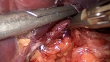
Dissection of the lower rectum is difficult in obese patients or in patients with a narrow and deep pelvis. It is in contact with vascular and nerve structures that must be preserved (Enker et al., 2000; Heald et al., 1998). To preserve genitourinary functions, care must be taken to avoid the middle hemorrhoidal vessels (which are occasionally large) and the parasympathetic nerve branches originating from posterior sacral foramina.
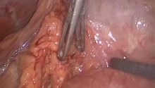
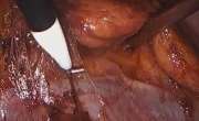
Dissection is performed posteriorly, laterally, then anteriorly to the rectum, alternating the right and left side as the rectum is progressively freed.
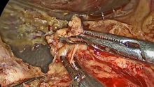
Laparoscopic freeing of the lower rectum is facilitated by the use of ultrasonic scissors or a vessel-sealing device.

[点击按纽展开更多->收起->回放]

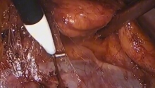
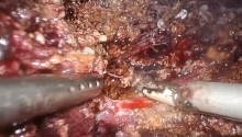
The posterior dissection is continued caudally on the posterior side of the rectum, anterior to the sacrum, once the sacrorectal ligament (Waldeyer’s fascia) has been opened. Care must be taken to avoid the anterior sacral venous plexus running on the anterior surface of the periosteum of the sacrum. This plexus is formed from posterior branches that lead to large 2-5 mm foramina from the third to the fifth sacral vertebrae before draining into the venous plexus of the intrasacral canal.
Further down the rectum is supported by the rectococcygeal ligament. Sharp division of this structure, using ultrasonic scissors or after coagulation, helps gain 1 to 2 cm over the posterior surface of the rectum, and also assists identification of the anal sphincter.

[点击按纽展开更多->收起->回放]

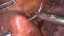
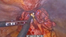
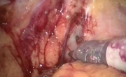
Dissection is facilitated by the posterior retraction of the rectum, and anterior retraction of the vagina or prostate with the use of an instrument introduced in the suprapubic trocar (trocar E), which grasps Denonvilliers’ fascia.
At this level, dissection of the anterior surface of the lower rectum can only be achieved after lateral rectal dissection (especially in men).
1. Prostate
2. Rectum

[点击按纽展开更多->收起->回放]

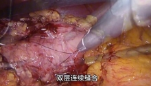
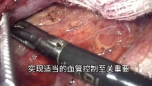
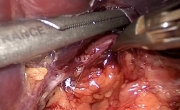
Low division of lateral ligaments exposes the lower pelvic space. This is done on the right side of the rectum, then on the left side. On the left side, the peritoneal reflection of the paracolic gutter should be incised down to the left lateral side of the rectum. One or more branches of the middle hemorrhoidal vessels joining the rectum on its anterior lateral surfaces are usually found on the lower part of lateral ligaments. These branches are infrequent, not always bilateral, and occasionally multiple. They should be controlled by clips or coagulation (bipolar forceps, vessel sealing device or ultrasonic scissors). At this level, the terminal branches of the pelvic nerve plexi run to the bladder, prostate, and genital organs (Enker et al., 1995; Enker et al., 2000).
 楼主| 发表于 2016-7-24 15:08:43 | 显示全部楼层
13. Splenic flexure
[点击按纽展开更多->收起->回放]

Mobilization of the splenic flexure is necessary to allow for a tension-free anastomosis in case of low colorectal or colo-anal anastomosis. The mobilization can take place at the beginning of the surgical procedure, before or after extraction of the rectum. The use of ultrasonic scissors is helpful, but not compulsory.
Except in a few patients with long and compliant sigmoid loops, mobilization of the left colon almost always mandates a large dissection, which combines division of lateral and posterior attachments with vascular divisions while preserving the vascular supply of the mobilized left colon. This vascular supply is sometimes difficult to assess laparoscopically.
1. Lateral approach
2. Medial approach

[点击按纽展开更多->收起->回放]

The medial mobilization of the splenic flexure is perfectly suited to the laparoscopic approach as the surgeon, situated to the patient’s right, has an excellent view of the anterior surface of the pancreas, the base of the left transverse mesocolon, and the posterior surface of the splenic flexure.
Mobilization is performed after the medial posterior freeing of the sigmoid mesocolon, which is then continued cephalad, anterior to Toldt’s fascia, then anterior to the pancreas.
1. IMV
2. Splenic vein
3. Base of the transverse mesocolon
4. Stomach

[点击按纽展开更多->收起->回放]

The lateral approach is conventional in open surgery. It is used in simple cases when the splenic flexure is mobile. It is completed with the freeing of the lateral and posterior attachments.
1. Attachments of omentum to the transverse colon
2. Phrenicocolic ligament
3. Toldt’s line
4. Toldt’s fascia
 楼主| 发表于 2016-7-24 15:08:50 | 显示全部楼层
14. Division/rectosigmoid
• Strategy
The division of the distal and proximal margins of resection is done prior to extraction. The extraction should be non-traumatic to avoid any risk of tumor cell dissemination (Okuda et al., 1998).
The specimen is isolated before extraction and placed in a plastic, watertight, tightly closed bag. The distal resection is usually performed first but if exposure is difficult, the proximal resection line may well be performed first (Okuda et al., 1998). The division should be done at least 2 cm below the inferior margin of the tumor and 10 cm above it, while removing the whole of the mesorectum.

[点击按纽展开更多->收起->回放]

Exclusion of lower rectum
The distal part of the dissected rectum is excluded either by ligature or by a clamp, or by a row of staples. The rectum distal to this exclusion is then washed with or without povidone iodine via the introduction of a cannula by the anal canal. This can help avoid the risk of tumor seeding on the division line. Lavage with an antiseptic and tumoricidal solution such as povidone iodine was proposed by Balli et al. (2000).

Rectal division
The lower rectum is exposed by gentle posterior traction. This traction can be performed with the thread used to ligate the rectum below the tumor to exclude the rectal stump. The division is performed with an articulated cutting linear stapler (45 mm, blue or green cartridges).

[点击按纽展开更多->收起->回放]

In rectal cancer, most surgeons perform the proximal division at the level of the sigmoid colon. The colon is divided on a portion that is healthy, supple, and well vascularized.
The division takes place on the descending colon if the sigmoid colon is affected by a concomitant pathology such as diverticulosis.
The division of the sigmoid mesocolon can be done either with a vessel sealing device, ultrasonic scissors or linear staplers. The complete sigmoid mesocolon is removed.
The division of the colon is done with a cutting linear stapler, which allows for clean division and stapling without opening the digestive tract.
The stapler (blue cartridges) is introduced in trocar C in the right iliac fossa.
1. Marginal arteries
2. Inferior mesenteric vessels

[点击按纽展开更多->收起->回放]

Once divided, the rectosigmoid is placed in a large, tightly closed plastic retrieval bag. The procedure can thus be continued without manipulation of the tumor in the abdominal cavity. The bag is introduced in trocar E or C. If the specimen is too bulky, it is extracted before completing mobilization of the left colon.
 楼主| 发表于 2016-7-24 15:08:57 | 显示全部楼层
15. Extraction
[点击按纽展开更多->收起->回放]

The extraction of the rectum is done through a mini-incision while protecting the abdominal wall with a wound protector. The specimen is isolated and placed into a tightly closed plastic bag before extraction. Certain authors recommend a thorough abdominal lavage after specimen extraction.

[点击按纽展开更多->收起->回放]

The incision is generally made in the suprapubic region. Other authors recommend the left lower quadrant. The ability to easily bring the proximal colonic resection line down to the left lower quadrant or to the right lower quadrant helps in the assessment of the mobilization of the colon into the pelvis and thus in the construction of a tension-free anastomosis.
The incision should be adapted to the size of the specimen to be extracted. Care must be taken not to crush the specimen at the time of the extraction.

[点击按纽展开更多->收起->回放]

The specimen is extracted in a tightly closed plastic bag. The abdominal incision is also protected with a wound protector (7 or 11 cm in diameter). This is also used to ensure that the abdominal cavity is airtight during the intracorporeal colorectal anastomosis that follows the extraction.

Wound protector
Once the wound protector is introduced into the abdominal opening, the plastic bag containing the resected sigmoid colon and rectum is brought up to the skin. The bag is then opened to extract the colon, while avoiding abdominal contamination.
1. Plastic bag
2. Plastic drape
 楼主| 发表于 2016-7-24 15:09:03 | 显示全部楼层
16. Direct anastomosis
[点击按纽展开更多->收起->回放]

The colorectal anastomosis is a delicate operative step. The rate of fistula, which can be higher in laparoscopic surgery, accounts for the difficulty of this step (Hartley et al., 2001). In our opinion, a diverting stoma to protect the low colorectal or colo-anal anastomosis is necessary, especially after radiation therapy.
A straight anastomosis can be performed, or after a colonic J-pouch has been fashioned.
The anastomosis includes both an extra-abdominal and an intra-abdominal step. The extra-abdominal step takes place after exteriorization of the left colon through the opening covered by the wound protector.

[点击按纽展开更多->收起->回放]

First, the colon is brought outside the abdominal cavity through the incision protected by the plastic drape. The colon should be easily exteriorized beyond the pubis, which helps evaluate the possibilities of lowering it to the pelvic floor for a tension-free anastomosis.
The vascular supply and compliance of the exteriorized colon are evaluated. With a spastic colon, it is recommended to fashion a J-shaped colonic pouch or to perform the anastomosis higher on the more compliant portion of descending colon. The omental appendages in the area of anastomosis are resected.
1. Pubic symphysis

[点击按纽展开更多->收起->回放]

The anvil of the circular stapler (at least 28 mm in diameter) is introduced once a purse-string suture has been fashioned on the distal part of the colon, either manually or with a purse-string device.
The anvil should then be easily introduced in the colonic lumen. Dilatation with bougies is unnecessary as it risks damaging the colonic wall. The quality of the compliance of the colon should allow for an effortless introduction of the anvil.
The purse is tightly closed around the shaft of the anvil. The left colon with the anvil is then pushed into the abdominal cavity.
1. 28 mm in diameter (at least)

[点击按纽展开更多->收起->回放]

The anastomosis can be performed under laparoscopic guidance after the reinsufflation of the pneumoperitoneum. The closure of the abdominal incision can be achieved by suturing the peritoneum and muscular planes. To ensure air seal, we prefer to close the previously placed wound protector, which gives the surgeon the possibility of exteriorizing the colon without reopening the abdominal wall.
This intra-abdominal step takes place entirely under laparoscopic guidance. Once the air seal of the abdominal cavity has been obtained with the closure of the plastic drape, the pneumoperitoneum is re-established. The pelvis is again perfectly exposed, with the help of a retractor that can be introduced through trocar D. Anastomosis is carried out with a mechanical circular stapler.
The circular stapler is introduced into the rectal stump by the assistant positioned between the patient’s legs, through the anus, which has previously been gently dilated. The rectal stump is then transfixed with the tip of the head of the circular stapler.
Once the anvil has been clicked onto the proximal part of the circular stapler, it is mandatory to confirm the absence of colon torsion. The posterior side of the mesocolon lies against the sacral concavity. The circular stapler is closed after ensuring no adjacent organs or epiploic appendages are trapped. Stapling is then done in keeping with the manufacturer's recommendations. Once the anastomosis is completed, the colon should mold into the sacral concavity.
The stapler is then loosened and removed through the anus.
 楼主| 发表于 2016-7-24 15:09:10 | 显示全部楼层
17. J-shaped anastomosis
[点击按纽展开更多->收起->回放]

Anastomosis can be performed on a colonic pouch. This mandates a better mobilization of the left colon, which is exteriorized through the protected opening. In case of suprapubic incision, the superior part of the pouch should be easily drawn beyond the pubis. The colonic J-pouch (6 to 7 cm high) is fashioned with linear staplers or sutures at the level of the antimesenteric tania. The distal end of the pouch is incised to introduce the anvil of the circular stapler.

[点击按纽展开更多->收起->回放]

The anvil of the circular stapler (at least 28 mm in diameter) is introduced after the fashioning of a purse-string suture at the distal end of the pouch. The purse-string is fashioned either manually or with a purse-string device.
The anvil should then be easily introduced into the colonic lumen. The purse is closed tightly around the shaft of the anvil. The left colon with the anvil is then pushed back into the abdominal cavity.

[点击按纽展开更多->收起->回放]

The anastomosis can be performed under laparoscopic guidance after the reinsufflation of the pneumoperitoneum. The closure of the abdominal incision can be achieved by suturing the peritoneum and muscular planes. To ensure air seal, we prefer to close the previously placed wound protector, which gives the surgeon the possibility of exteriorizing the colon without reopening the abdominal wall.

The anvil, which has been placed at the apex of the pouch, is drawn into the pelvis. It is then clicked onto the proximal part of the circular stapler. The pouch is positioned anteriorly, as its mesentery is situated in contact with the sacral concavity. Once the anastomosis has been completed, the colon and rectum should mold into the sacral concavity. Because of the straight trajectory of the colon, there is not only an increased risk of leakage, but also a risk of secondary fecal incontinence, due to the absence of the natural anorectal angle (90° at rest).
 楼主| 发表于 2016-7-24 15:09:17 | 显示全部楼层
18. End of the procedure
[点击按纽展开更多->收起->回放]

Verification of the anastomosis is mandatory. This includes checking for the circular aspect of the amputated rectal and colon rings, optional air test, and for some authors (Franklin et al., 1996) endoscopic transanal evaluation of the anastomosis. An air test is almost impossible to perform in very low anastomoses.

Air test:
The pelvis is filled with a saline solution so that the anastomosis is largely covered. One clamp is positioned on the colon at the level of the promontory. Air is then injected under low pressure with a syringe introduced in the anal canal. The absence of bubbles confirms the absence of leakage of the anastomosis, but does not mean there is no risk of postoperative fistulas.

Dye test:
The dye test completes the air test if it is positive. Once the pelvis is clean, a betadine solution is injected under low pressure into the rectum with a syringe. This helps identify leaks.
If there is a minimal anterior leak, a suture can be performed. In case of massive leak, the anastomosis should be repeated either transanally, or via the abdominal route (laparoscopy or open surgery). In such cases, a diverting stoma (preferably ileostomy) is often necessary.
Methylene blue test can replace the betadine test. The problem is that it stains the pelvis, which hinders continuation of the exploration. We do not use it.
Drainage of the abdominopelvic cavity is not routine. It is done only if persistent oozing occurs.
1. Air

[点击按纽展开更多->收起->回放]

A protective stoma is recommended in low colorectal anastomosis, especially after radiation therapy because of the higher risk of fistula (Heald et al., 1998). It reduces the clinical severity of potential fistulas, which has a rate of 15% in our experience. The loop ileostomy is favored by most authors. It is situated preferably on the terminal ileum to better preserve the vascular supply of the anastomosis. It can also be situated on the right transverse colon. The selected bowel segment is identified under laparoscopic guidance before it is drawn to the skin.
1. Ileostomy
2. Transverse colostomy

• Closure
We conduct moderate lavage of the abdominal cavity before and after the anastomosis.
Exsufflation of the abdominal cavity is recommended before trocar extraction. The risk of incarceration of the omentum or the small bowel is avoided. Tumor seeding can also be avoided. For the same reason, irrigation of trocar incisions with a betadine solution is recommended by some authors (Balli et al., 2000).
The trocar incisions are closed cautiously. Incisions larger than 5 mm should be closed in layers to prevent incisional hernias. In obese patients, instruments such as the traumatic French needle can be very useful.
The gastric tube is not routinely left in place. Many surgeons remove it as soon as the patient awakes. We remove it on POD1.
The patient is mobilized as soon as possible on the operative day. Food intake is resumed as soon as bowel functions are restored. The urinary catheter should be rapidly removed to avoid any infection. Prevention of deep vein thrombosis is routinely performed (low molecular weight heparin).
 楼主| 发表于 2016-7-24 15:09:25 | 显示全部楼层
19. Conclusions
Total mesorectal excision is the total, “en bloc” resection of the rectum and perirectal lymph node tissue (mesorectum) from the pelvic inlet to the pelvic floor. However, this resection presents risks such as hemorrhage, nerve injuries, local recurrences, fistulas, and anastomotic stenoses (Hartley et al., 2001).
The magnification of anatomical structures during pelvic dissection and perfect visualization of the procedure favor the teaching and broadcasting of the surgical technique.
Even though laparoscopy has not demonstrated evidence of better oncological efficacy in the long run, initial results are promising. Our experience shows that this procedure is performed via laparoscopy with similar results to the ones obtained in open surgery. Better preservation of immunity (Nishiguchi et al., 2001) can allow for immediate administration of adjuvant therapy in node positive patients, which will hopefully lead to improved survival outcomes in the future.
Learning should be provided with the help of a surgical team skilled in colorectal and laparoscopic surgery.
您需要登录后才可以回帖 登录 | 注册

本版积分规则

丁香叶与你快乐分享

微信公众号

管理员微信

服务时间:8:30-21:30

站长微信/QQ

← 微信/微信群

← QQ

Copyright © 2013-2024 丁香叶 Powered by dxye.com  手机版 
快速回复 返回列表 返回顶部