训练用单针/双针带线【出售】-->外科训练模块总目录
0.5、1、2、3.5、5mm仿生血管仿生体 - 胸腹一体式腹腔镜模拟训练器
仿气腹/半球形腹腔镜模拟训练器
[单端多孔折叠]腹腔镜模拟训练器
「训练教具器械汇总」管理员微信/QQ12087382[问题反馈]
开启左侧

[资源] 腹腔镜下乙状结肠憩室炎(图文演示)

[复制链接]
 楼主| 发表于 2016-7-26 09:24:06 | 显示全部楼层
10. Post medial dissection
[点击按纽展开更多->收起->回放]

Mobilization of the colon follows vascular division. It may be performed medially or laterally. We prefer a medial approach, which is well adapted for laparoscopy, as it preserves the working space and demands the least handling of the sigmoid colon. If a fixed colon is encountered, the surgeon must know how to alternate between medial and lateral approaches according to the difficulties encountered.

[点击按纽展开更多->收起->回放]

The objective of this step is to expose the anterior surface of Toldt's fascia. This fascia protects the retroperitoneal structures.

[点击按纽展开更多->收起->回放]

After division of the vessels, the mesosigmoid is retracted anteriorly (trocar E) to open the posterior space.

[点击按纽展开更多->收起->回放]

1. Ureter
2. Genital vessels
3. Posterior surface of mesosigmoid
4. Lateral layer of mesosigmoid
5. Mesorectum
The dissection plane is situated between Toldt's fascia and the mesosigmoid. This plane is avascular and easily dissected. It is exposed in the course of the dissection along the posterior surface of the mesosigmoid.
Once Toldt's fascia has been exposed, the detachment of the mesosigmoid is pursued posteriorly and laterally to the white line of Toldt.
The left sympathetic nerve trunk, the ureter, and genital vessels, covered by Toldt's fascia, are viewed during the dissection.
 楼主| 发表于 2016-7-26 09:24:14 | 显示全部楼层
11. Resection/mesorectum
[点击按纽展开更多->收起->回放]

We recommend a partial resection to preserve the superior rectal vessels.

[点击按纽展开更多->收起->回放]

We recommend partial resection of the mesorectum, anterior to the superior rectal vessels, to preserve the vascular supply to the rectum. This is performed above the origin of the first superior rectal branch, which is divided. This branch is an excellent landmark for the posterior surface of the colorectal junction. The division of the mesorectum is thereafter avascular.
 楼主| 发表于 2016-7-26 09:24:41 | 显示全部楼层
12. Lateral mobilization
[点击按纽展开更多->收起->回放]

The sigmoid loop is pulled toward the right hypochondrium (grasper in trocar F) to exert traction on the left layer of the mesosigmoid.

[点击按纽展开更多->收起->回放]

The white line of Toldt is opened cephalad and caudally.
Laterally, care must be taken to avoid the genital vessels and the left ureter. Identification of the ureter with a ureteral catheter is usually not necessary. When difficulty is encountered in identifying the ureters, it is preferable to pursue its dissection higher up in a non-inflamed area, using a medial approach, and to follow it caudally into the operative field.
After incision of the left layer of the mesosigmoid, the lateral dissection joins the plane of the previously performed posterior dissection along Toldt's fascia.

[点击按纽展开更多->收起->回放]

1. Sigmoid colon
2. Mesosigmoid
3. Adhesions
In the case of inflammatory adhesions of the sigmoid colon to the abdominal wall, lateral mobilization can be difficult. In this case, it is advisable to perform a medial posterior approach to identify the ureter in a healthy, non-inflamed area.
 楼主| 发表于 2016-7-26 09:24:55 | 显示全部楼层
13. Division/sigmoid
[点击按纽展开更多->收起->回放]

Once it is freed, the sigmoid colon is resected. Total resection of the sigmoid colon, including the rectosigmoid junction, is performed. Cephalad, the proximal division is performed on a portion of the colon that is supple and without any diverticula.

[点击按纽展开更多->收起->回放]

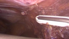
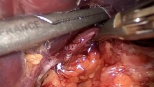
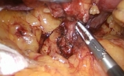
After division of the mesorectum, the distal division of the sigmoid colon is performed below the rectosigmoid junction. The junction is identified on the anterior surface of the rectum by the coalescence of the anterior tenia, and posteriorly by the first collateral branch of the superior rectal vessels.
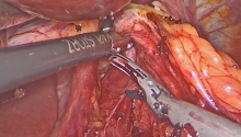
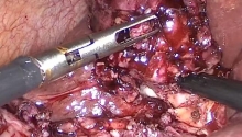
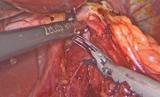
Division of the rectum is performed using a mechanical linear stapling device, which can divide and staple without opening the digestive tract. The stapler is introduced through trocar C into the right iliac fossa. We prefer using staples for thick tissues (green cartridges, 4.8 mm), applied perpendicular to the digestive tract. The use of reticulating staplers may be useful.

• Proximal division
[点击按纽展开更多->收起->回放]

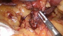
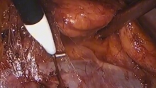
The proximal division should be performed in a non-inflamed area on a perfectly healthy and supple portion of the colon. It includes division of the mesocolon, followed by division of the colon. The division of the mesocolon is always performed in the abdominal cavity. The colon may be divided outside of the abdominal cavity, after it is exteriorized.

[点击按纽展开更多->收起->回放]

Division is performed with a high-frequency hemostasis device, ultrasonic dissectors, monopolar scissors or linear staplers. The marginal arteries are preserved up to the area of transection.
Whether the colon is divided intracorporeally or extracorporeally, it is important to perform this mesocolic division first because it prevents the mesentery from tearing (during the extraction of the sigmoid colon).

[点击按纽展开更多->收起->回放]

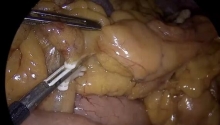
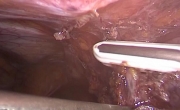
Intracorporeal division is used in surgery for cancer of the sigmoid colon. It is usually performed with a mechanical stapler (blue cartridges) introduced through trocar C into the right iliac fossa. The sigmoid colon is then completely freed.

[点击按纽展开更多->收起->回放]

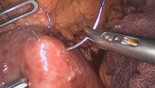
In sigmoid colectomy for diverticulis, the colon is usually exteriorized before it is divided. The exteriorization is pursued until a healthy portion of colon is reached. The preceding division of the mesocolon facilitates this.
 楼主| 发表于 2016-7-26 09:25:18 | 显示全部楼层
[点击按纽展开更多->收起->回放]

In the event that a long segment of sigmoid colon is resected, mobilization of the left colon is necessary to allow for a tension-free colorectal anastomosis.
The left colon is freed by division of its posterior and lateral attachments. Occasionally, division of the left colic vessels is required for full mobilization of the left colon.
The vascular supply of the mobilized left colon should be preserved. This is often difficult to assess in laparoscopic surgery.
Mobilization of the splenic flexure is frequently required. This can be achieved in a number of different ways. It is important for the surgeon to be familiar with all these approaches in order to select the approach most suitable for each case.
 楼主| 发表于 2016-7-26 09:25:32 | 显示全部楼层
15. Lateral mobilization
[点击按纽展开更多->收起->回放]

1. Attachments of omentum to colon
2. Phrenicocolic ligament
3. Toldt's line
4. Toldt's fascia
This approach is conventional in open surgery. It is used in simple cases with an easily mobilized splenic flexure. It includes freeing of the lateral and posterior attachments.

[点击按纽展开更多->收起->回放]

An ascending incision is made along the white line of Toldt using scissors introduced via trocar D.

[点击按纽展开更多->收起->回放]

The phrenocolic ligament is then divided using scissors introduced through trocar D. Retracting the colon and the splenic flexure toward the right iliac fossa using graspers introduced through trocars C and E helps in this exposure.

[点击按纽展开更多->收起->回放]

Attachments are incised close to the colon until the omental bursa (lesser sac) is opened. Division of these attachments is pursued as needed toward the right, to facilitate lowering of the left transverse colon.
 楼主| 发表于 2016-7-26 09:25:45 | 显示全部楼层
16. Medial mobilization
[点击按纽展开更多->收起->回放]

Medial mobilization is perfectly suited to the laparoscopic approach as the surgeon, situated to the patient's right, has an excellent view of the anterior surface of the pancreas and the base of the left transverse mesocolon.
It follows the medial posterior freeing of the mesosigmoid, which then continues upward, anterior to Toldt's fascia.

[点击按纽展开更多->收起->回放]

The splenic flexure is freed on its posterior surface while remaining close to the left mesocolon. This avoids the risk of a dissection posterior to the pancreas with an attendant injury of the splenic vein. Division of the IMV just below the inferior border of the pancreas facilitates this operative step.

[点击按纽展开更多->收起->回放]

On the anterior inferior border of the pancreas, the base of the transverse mesocolon is identified. This is divided caudad to cephalad and from right to left, opening the omental bursa (lesser sac). Anterior to the pancreatic tail, care must be taken not to injure the pancreas and small mesocolic vessels, which course through the left transverse mesocolon.
 楼主| 发表于 2016-7-26 09:25:56 | 显示全部楼层
17. Extraction
[点击按纽展开更多->收起->回放]

Extraction is performed through a mini-incision while protecting the abdominal wall.

• Incision
[点击按纽展开更多->收起->回放]

The size of the specimen, its location, and the extraction technique take into account the volume of the specimen, the patient’s body habitus, and cosmetic concerns.

[点击按纽展开更多->收起->回放]

The incision should be adapted to the size of tissues to be extracted. Care must be taken not to crush the specimen during extraction.

[点击按纽展开更多->收起->回放]

The incision is generally performed in the suprapubic region (1), more rarely in the right iliac fossa (2), but never in the left iliac fossa (3). The lowering of the colon in (1) or (2) allows evaluation of the possibilities of mobilization of the colon in the pelvis. If the mobilized colon reaches the extraction area in a tension-free manner, it may be safely assumed the anastomosis will be tension free as well.

[点击按纽展开更多->收起->回放]

The wall should be protected to avoid bacterial and cellular contamination (risk of wound implantation of cancer cells). This protection is made up of a waterproof plastic-coated drape with a ring (7 or 11 mm in diameter).
The same plastic-coated protection is used during intracorporeal anastomosis. The bag is twisted on itself to reseal the peritoneal cavity in a reversible fashion.

• Extraction
[点击按纽展开更多->收起->回放]

The extraction of the specimen is performed either using an airtight plastic bag or through a plastic-coated drape with a ring that allows reestablishment of the pneumoperitoneum following the extraction of the specimen.
In our current practice, we frequently combine the techniques.

[点击按纽展开更多->收起->回放]

The resected sigmoid colon is extracted through the plastic drape introduced into the abdominal incision.
The intra-abdominal division of the mesocolon prevents it from tearing during extraction, especially in obese patients.

• In a plastic-coated bag
 楼主| 发表于 2016-7-26 09:26:16 | 显示全部楼层
18. Preparation/anastomosis
[点击按纽展开更多->收起->回放]

The colorectal anastomosis is a delicate step. We always use a mechanical circular stapling device to transfix the rectal stump.
Performing the anastomosis includes an extra-abdominal preparatory step and an intra-abdominal step performed laparoscopically
The extra-abdominal step takes place after exteriorization of the left colon through the incision protected by the plastic-coated drape.

[点击按纽展开更多->收起->回放]

If necessary, the colon is cut again in a healthy, supple and well-vascularized zone. The anvil (at least 28 mm in diameter) is then introduced into the colonic lumen and closed with a purse string. The left colon with the anvil is then reintroduced into the abdominal cavity.

[点击按纽展开更多->收起->回放]

The closure ensures the airtightness of the abdominal cavity required to perform the anastomosis under laparoscopic guidance. The closure is achieved either by primary closure of the abdominal wall or by introducing a plastic-coated drape with an airtight ring. The second option is more attractive because it leaves the surgeon the possibility of exteriorizing the colon if necessary.
 楼主| 发表于 2016-7-26 09:26:25 | 显示全部楼层
19. Anastomosis
[点击按纽展开更多->收起->回放]

The intra-abdominal step is performed entirely under laparoscopic guidance after reinflation of the abdominal cavity. It includes transfixing the rectal stump and performing the colorectal anastomosis with a circular mechanical stapler.

[点击按纽展开更多->收起->回放]

After ensuring that the abdominal cavity is airtight, the pneumoperitoneum is reestablished. The circular stapler is introduced into the rectum through the atraumatically dilated anus. The rectal stump is then transfixed with the tip of the head of the circular stapler.
To avoid ischemic risks, the stapler can be introduced in the middle or at one of the ends of the rectal stump staple line while avoiding leaving lateral ear-shaped flaps.
In women, the posterior vaginal wall should be retracted anteriorly.

[点击按纽展开更多->收起->回放]

Once the anvil has been clicked onto the proximal part of the circular stapler, it is mandatory to ensure that the proximal part of the colon is not twisted. The stapler is then closed; the surgeon should check that no neighboring organs are incarcerated before stapling in accordance with the manufacturer's recommendations.
The stapler is then loosened and withdrawn through the anus.

[点击按纽展开更多->收起->回放]

Verification of the anastomosis is mandatory. This includes checking for the circular aspect of the amputated rectal and colon rings, optional air test and for some authors, endoscopic transanal evaluation of the anastomosis.
您需要登录后才可以回帖 登录 | 注册

本版积分规则

丁香叶与你快乐分享

微信公众号

管理员微信

服务时间:8:30-21:30

站长微信/QQ

← 微信/微信群

← QQ

Copyright © 2013-2024 丁香叶 Powered by dxye.com  手机版 
快速回复 返回列表 返回顶部