训练用单针/双针带线【出售】-->外科训练模块总目录
0.5、1、2、3.5、5mm仿生血管仿生体 - 胸腹一体式腹腔镜模拟训练器
仿气腹/半球形腹腔镜模拟训练器
[单端多孔折叠]腹腔镜模拟训练器
「训练教具器械汇总」管理员微信/QQ12087382[问题反馈]
开启左侧

[资源] 腹主动脉瘤:经腹腔内镜手术(图文演示)

[复制链接]
 楼主| 发表于 2016-7-29 12:56:47 | 显示全部楼层
10. Dissection II
[点击按纽展开更多->收起->回放]

1. Infrarenal aortic aneurysm
2. IMA
3. Division of the IMA
Often caught in the periaortic inflammation, the trunk of the IMA may be difficult to free because of the presence of an occasionally hemorrhagic venous plexus. It is helpful to use an atraumatic, curved vessel retractor for this step.
If preoperative studies indicate thrombosis, or if it can be occluded without danger, the IMA is divided with high-frequency forceps or between 2 clips. In all other cases, it is preferable to examine the IMA and check its flow after opening the aortic aneurysm to decide whether a reimplantation is required.

[点击按纽展开更多->收起->回放]

1. Right and left common iliac arteries
2. Bifurcation
Dissection of the IMA generally frees the retroperitoneal attachments that interfere with aortic dissection. It is then easier to reach the bifurcation by incising the median peritoneum anterior to the promontory. The anterior surface of the aorta is progressively exposed, followed by exposure of the left common iliac artery, which can be entirely freed in certain patients, though only partially in others. The first few centimeters of the right common iliac artery are then dissected.
While freeing the common iliac arteries, the dissection of their postero-internal surfaces involves a risk of vein injury, especially to the common iliac veins and their branches. This part of the dissection must be done extremely carefully, using atraumatic instruments (curved vessel retractor). It can be avoided if aorto-aortic restoration is done with a straight graft, in which case a simple clamping of the lateral surfaces of the common iliac arteries may be sufficient.

[点击按纽展开更多->收起->回放]

1. Clipped left lumbar artery
At this point in the procedure, only the left lumbar arteries can be freed and occluded using clips. In certain patients, this dissection is made difficult by the fatty and lymphatic tissue that surrounds these vessels and that is often hemorrhagic.

• Right aortic surface
This final step of the dissection should not be performed until the anterior surface and left side of the aorta have been sufficiently freed.
The anterior surface of the aneurysm can interfere with the dissection of the right side. It is therefore advisable to place the operating trocars and the optical trocar D closer to the midline in case of a large aneurysm with anterior protrusion.
The main risk here is to the inferior vena cava. As in conventional surgery, this risk is avoided by performing the dissection against the aortic wall.
Once the aortic-iliac dissection has been achieved, it easier to reach the aortic neck and to free its posterior surface if necessary. Often the occlusion of the right lumbar arteries with clips requires preliminary proximal aortic clamping.
 楼主| 发表于 2016-7-29 12:56:54 | 显示全部楼层
11. Clamping
• Principle
While is preferable to introduce the proximal aortic clamp laparoscopically, the iliac clamps are often easier to introduce via minilaparotomy.

[点击按纽展开更多->收起->回放]

Two types of proximal laparoscopic aortic clamps may be used:
1. Straight aortic clamp
2. Reverse angled aortic clamp

[点击按纽展开更多->收起->回放]

1. Straight aortic clamp
The aortic clamp is introduced laparoscopically through a 10 mm longitudinal incision, placed at the level of the neck of the aneurysm, without using a trocar. Before making this incision, in order to make sure that it is correctly placed, it is advisable to insert an intramuscular needle and to check its point of projection on the aorta with the laparoscope.
While introducing the clamp, it is important to verify that the hinge between its jaws has completely entered the peritoneal cavity before opening it. Depending on the size of the patient and of the aorta, a long or short aortic clamp may be used. When opening the clamp, the surgeon should use the laparoscope to make sure that the jaws are correctly positioned laterally and that the neck of the aorta is fully clamped.

[点击按纽展开更多->收起->回放]

1. Reverse angled aortic clamp
This clamp is particularly useful if the neck of the aorta angles towards the left, or if it seems that it will be difficult to sew the back wall.
It is introduced through the incision of trocar C after removing the trocar. The penetration of the tip of the clamp to the hinge of its jaws is achieved under laparoscopic guidance. The clamp is then opened and the inferior jaw is placed on the posterior surface of the aortic neck.
 楼主| 发表于 2016-7-29 12:57:00 | 显示全部楼层
12. Minilaparotomy
[点击按纽展开更多->收起->回放]

After stopping the insufflation, all trocars are removed. Only the laparoscopic intestinal retractor is left in place. A 6 to 9 cm median minilaparotomy, which is usually supraumbilical, is then performed. The incision is begun at the trocar A incision. The minilaparotomy should take several factors into consideration:
If an aortic tube is used, the minilaparotomy should be centered on the mid-portion of the aneurysm to perform the 2 aortic anastomoses by alternating traction on the wall (upwards and then downwards).
If an aorto-bifemoral bypass is performed, the minilaparotomy is shifted slightly towards the proximal aortic neck.
If an aorto-iliac or aorto-ilio-femoral bypass is performed, the minilaparotomy must be longer (9 to 10 cm).

• Body habitus of patient
In obese patients, especially in those with thick mesenteries, a longer minilaparotomy incision is required.

• Suturing difficulties
For various reasons (aorta with inflammation or with a fragile wall, periaortic adhesions) it may be necessary to enlarge the minilaparotomy, usually upwards. Experience has shown that the postoperative course depends very little on the length of the minilaparotomy, especially when compared to the risks related to a poorly performed aorto-prosthetic anastomosis.

[点击按纽展开更多->收起->回放]

1. Suction cannula
The surgeon may remain on the patient’s left or move to the patient’s right, opposite the first assistant. A 6 to 9 cm cutaneous incision is made, which may be slightly longer on the underlying adipose and aponeurotic planes.
As soon as the CO2 has been evacuated from the peritoneal cavity, it is possible to connect the cell saver to the end of the suction cannula. At the same time, a normal dose of an intravenous bolus injection of heparin is administered by the anesthesiologist.
The remainder of the procedure is performed under the surgeon’s direct vision in the abdominal cavity. The laparoscopic retractor for the intestinal loops enables the surgeon to view the abdominal aortic aneurysm that has just been dissected. Self-retaining retractors are placed on each side of the incision and maintain exposure.
 楼主| 发表于 2016-7-29 12:57:08 | 显示全部楼层
[点击按纽展开更多->收起->回放]

The proximal aortic clamp is closed using the security notch. The bulldog clamps are applied on the common iliac arteries. The aneurysm is first opened longitudinally, and then on the right and left lateral surfaces on both ends of the incision. The mural thrombus is removed and sent for bacteriologic culture. It is then possible, via an endo-aneurysmal approach, to occlude the lumbar arteries (the right, in particular) that have a backflow. This is achieved with a non-absorbable figure-eight suture. This step may need to be repeated on the median sacral artery.
If there is doubt concerning the advantage of a direct reimplantation of the IMA, the bulldog clamp can be released for a few instants to evaluate the quality of the backflow.
 楼主| 发表于 2016-7-29 12:57:17 | 显示全部楼层
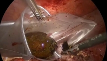
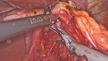
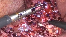
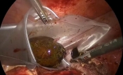
14. Graft implantation
[点击按纽展开更多->收起->回放]

1. Proximal aortic cuff
2. Opened aneurysm
3. Aortic graft
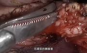
4. Running suture being performed, with medial to lateral insertion of the needle in the aorta
5. Biological glue
A polyester or polytetrafluoroethylen graft is chosen that is adapted to the diameter of the aortic neck and the type of bypass being done (aortic tube or bifurcated graft).
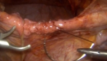
The body of the graft is cut to form a slightly oblique slant. Double-threaded 3.0 or 4.0 vascular suture, 1.2 m in length, is used.
The surgeon performs a conventional end-to-end anastomosis of the graft to the aorta while the first assistant maintains the suture under tension. The seal of this anastomosis is tested at the end of the procedure and biological glue is often added.

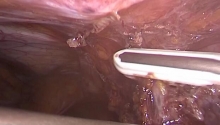
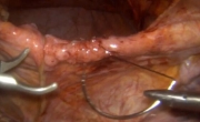
• Distal anastomoses
[点击按纽展开更多->收起->回放]

It is often necessary to reapply the self-retaining retractors to exert a slight downward traction on the wall. Calcifications and plaques may mandate modification of the distal anastomosis.
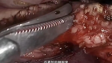
Two bulldog clamps were previously applied to the origin of the common iliac arteries. The distal part of the straight graft is cut with a slight slant to the desired length and a second, conventional anastomosis of the graft to the aorta is performed following the method described above.

[点击按纽展开更多->收起->回放]

Aorto-iliac bypass: when the aneurysm involves the bifurcation, distal anastomoses on the first few centimeters of the common iliac arteries are performed, above the previously applied bulldog clamps. The origin of each iliac artery is opened longitudinally and transversally. Each branch of the bifurcated graft is cut with a slant to the desired length.
A conventional end-to-end anastomosis on the graft to the common iliac artery is performed on each side, using 4.0 or 5.0 vascular suture.

[点击按纽展开更多->收起->回放]

Aorto-bifemoral bypass: involvement of the aneurysm down to the common iliac arteries requires an extension of the bypass to the common femoral arteries, or occasionally to the external iliac arteries.
In these cases, a classical approach via the right and left femoral triangles is used at the beginning of the procedure, before the abdominal approach. During this approach, and to avoid a secondary CO2 leak through the inguinal incisions, the arterial dissection below the crural arch must not be pursued.
During the abdominal approach, the common iliac arteries may be occluded with sutures or a linear stapler. Each branch of the graft must be placed in a subperitoneal anatomical position. A standard curved aortic clamp is introduced through the femoral triangle towards the aortic area. To guide the clamp during its insertion, the surgeon places his or her hand in the abdomen through the minilaparotomy.
Once each branch of the graft is exteriorized at the level of the femoral triangles, a conventional femoral-graft anastomosis is carried out with a 5.0 or 6.0 vascular suture. This anastomosis is done in an end-to-side fashion, permitting retrograde perfusion of the internal iliac arteries via the external iliac arteries.

[点击按纽展开更多->收起->回放]

Aorto-ilio-femoral bypass: if the aneurysm extends down asymmetrically, requiring a bypass onto one of the femoral axes, a limb of the graft is anastomosed to a common iliac artery. The other limb is passed into a retroperitoneal anatomical position and laterally anastomosed to the femoral bifurcation.

• Special cases
[点击按纽展开更多->收起->回放]

Bypass onto the iliac bifurcations: in the specific case where one or both internal iliac arteries must be perfused in an anterograde way (abdominal aortic aneurysm extending onto the origin of one or both internal iliac arteries) it is necessary to perform one or two anastomoses of the grafts to the iliac bifurcation(s).
On the left side, the laparoscopic dissection, which can be extended by an enlarged downward minilaparotomy, usually permits the surgeon to reach the iliac bifurcation and to perform the graft to iliac anastomosis at this level.
On the right side, laparoscopic dissection of the iliac bifurcation is difficult. A complementary 5 to 7 cm cutaneous incision (in the right flank) is usually preferable. The right iliac bifurcation can be reached by a retroperitoneal approach through this incision. The right limb of the graft can be easily placed in anatomical position.

• Case 2
Combined aorto-ilio-femoral occlusive lesions: the association of severe occlusive lesions with an abdominal aortic aneurysm is not frequent. It should be suspected in the presence of symptoms involving the legs (intermittent claudication, ischemic pain at rest or ischemic trophic disorders, disappearance of pulse). Systematic preoperative CT scan with contrast can often reveal occlusive lesions. In the presence of these signs, aorto-arteriography of the legs is mandatory.
In most cases, the type of bypass performed is an aorto-bifemoral restoration that takes this information into account.

[点击按纽展开更多->收起->回放]

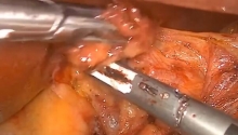
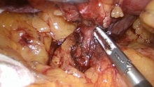
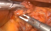
1. Left inferior polar renal artery
2. IMA
Reimplantation of the IMA: the decision to reimplant the IMA into the left surface of the body of the graft or into the left branch of the bifurcated aortic graft can be made either before the procedure following the preoperative workup (occlusion of one or both internal iliac arteries) or if weak backflow is discovered when releasing the clamp from the IMA after opening the abdominal aortic aneurysm.
As in conventional surgery, an aortic patch is cut around the IMA ostium and reimplantation is performed through the minilaparotomy with a running 5.0 or 6.0 vascular suture.
 楼主| 发表于 2016-7-29 12:57:24 | 显示全部楼层
15. Closure
• Closure
After placement of the graft and checking hemostasis, a retroperitoneal closure can be performed through the minilaparotomy, using slowly absorbable suture.

• Removal of the retractor
The scrub nurse should count the sponges to make sure that they have all been removed from the abdomen. The laparoscopic bowel retractor is then dismantled. The traction sutures should first be cut outside the abdomen and their internal portion (plastified cuff) recuperated. The metal arm of the bowel retractor is then detached from the operating table. The polypropylene mesh is removed through the minilaparotomy. The metal arm is slipped out of the abdomen through the opening into which it was inserted.

[点击按纽展开更多->收起->回放]

The minilaparotomy and the trocar wounds are closed in 2 or 3 planes, in a conventional fashion without drainage. If necessary, the femoral triangles are closed in 3 planes over a drain.
 楼主| 发表于 2016-7-29 12:57:42 | 显示全部楼层
16. Conclusion
Video-assisted endoaneurysmorrhaphy for abdominal aortic aneurysms can be used to obtain the same results as in conventional surgery, with a less aggressive abdominal approach.
Early results have shown a decrease in postoperative pain and in the risk of nosocomial infection. Bowel function is rapidly restored and the patient can usually be discharged from the hospital between POD3 and POD6. In our experience, no delayed reoperations of the abdominal area were necessary (mean follow-up: 22 months, 3-36 month range) (Alimi et al., 2001).
Other authors (Edoga et al., 1998) have described a retroperitoneal laparoscopic approach with encouraging results. For surgeons who are not yet experienced in the technique, a transperitoneal approach using the “less dominant” hand introduced through the minilaparotomy, while maintaining a pneumoperitoneum, can be a reassuring option (Kolvenbach et al., 2001).
In the future, the development of new instruments will make it possible to decrease the operative time and the duration of the clamping, which are still slightly longer than for conventional surgery. It is probable that it will even be possible to avoid the minilaparotomy in certain patients.
The smooth performance of video-assisted endoaneurysmorrhaphy for abdominal aortic aneurysms depends on:
- the quality of the equipment;
- adherence to the operative strategy, which is the same as that used in conventional surgery;
- the experience of the surgical team in laparoscopic techniques.
After training on animals or cadavers, these techniques should be practiced with a highly skilled team. Comparative clinical studies of good methodological quality, involving a sufficient number of patients, will be needed to assess this technique.
 楼主| 发表于 2016-7-29 12:57:54 | 显示全部楼层
17. Reference
Alimi YS, Hartung O, Valerio N, Juhan C. Laparoscopic aortoiliac surgery for aneurysm and occlusive
disease: when should a minilaparotomy be performed? J Vasc Surg 2001;33:469-75.
Creech O, Jr. Endo-aneurysmorrhaphy and treatment of aortic aneurysm. Ann Surg 1966;164:935-46.
Edoga JK, Asgarian K, Singh D, James KV, Romanelli J, Merchant S et al. Laparoscopic surgery for
abdominal aortic aneurysms. Technical elements of the procedure and a preliminary report of the first
22 patients. Surg Endosc 1998;12:1064-72.
Kolvenbach R, Ceshire N, Pinter L, Da Silva L, Deling O, Kasper AS. Laparoscopy-assisted aneurysm
resection as a minimal invasive alternative in patients unsuitable for endovascular surgery. J Vasc
Surg 2001;34:216-21.
您需要登录后才可以回帖 登录 | 注册

本版积分规则

丁香叶与你快乐分享

微信公众号

管理员微信

服务时间:8:30-21:30

站长微信/QQ

← 微信/微信群

← QQ

Copyright © 2013-2024 丁香叶 Powered by dxye.com  手机版 
快速回复 返回列表 返回顶部