15. Reference
Azorin J, Lamour A, Destable MD, de Saint-Florent G. La péricardoscopie: définition, intérêt et limite. Rev
Pneumol Clin 1986;42:138-41.
Gossot D, Mourey F, Roland E, Celerier M. Abord thoracoscopique des épanchements péricardiques.
Presse Med 1994;23:1480-2.
Hazelrigg SR, Mack MJ, Landreneau RJ, Acuff TE, Seifert PE, Auer JE. Thoracoscopic pericardiectomy
for effusive pericardial disease. Ann Thorac Surg 1993;56:792-5.
Krasna M, Fiocco M. Thoracoscopic pericardiectomy. Surg Laparosc Endosc 1995;5:202-4.
Nakamoto H, Suzuki T, Sugahara S, Okada H, Kaneko K, Suzuki H. Successful use of thoracoscopic
pericardiectomy in elderly patients with massive pericardial effusion caused by uremic pericarditis. Am J
Kidney Dis 2001;37:1294-8.
Ohtsuka T, Wolf RK, Wurnig P, Park SE. Thoracoscopic limited pericardial resection with an ultrasonic
scalpel. Ann Thorac Surg 1998;65:855-6.
Urschel JD, Horan TA. Pericardioscopy and biopsy. Surg Endosc 1993;7:100-1.
Wurtz A, Chambon JP, Millaire A, Saudemont A, Ducloux G. La péricardoscopie : techniques, indications
et résultats. A propos d'une expérience de soixante-dix cas. Ann Chir 1992;46:188-93. |

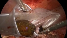
 急性胆囊炎——紧急救治策略
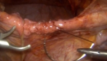
 采用腹腔镜经筋膜腹外缝合术修复因 Morgagn
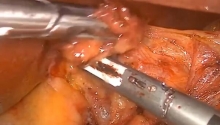
 腹腔镜全胃切除术治疗胃癌中采用线性吻合器
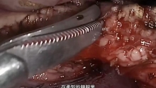
 腹腔镜下Whipple手术治疗胰头肿瘤

 楼主
楼主
 显身卡
显身卡