16. Reference
Bruhat MA, Manhes H, Mage G, Pouly JL. Treatment of ectopic pregnancy by means of laparoscopy.
Fertil Steril 1980;33:411-4.
Dubuisson JB, Morice P, Chapron C, De Gayffier A, Mouelhi T. Salpingectomy - the laparoscopic
surgical choice for ectopic pregnancy. Hum Reprod 1996;11:1199-203.
Philippe E, Ritter J, Lefakis P, Laedlein-Greilsammer D, Itten S, Foussereau S. Grossesse tubaire,
ovulation tardive et anomalie de nidation. Gynecol Obstet (Paris) 1970;69:617+.
Pouly JL, Chapron C, Manhes H, Canis M, Wattiez A, Bruhat MA. Multifactorial analysis of fertility after
conservative laparoscopic treatment of ectopic pregnancy in a series of 223 patients. Fertil Steril
1991;56:453-60.
Pouly JL, Mage G, Gachon F, Gaillard G, Bruhat MA. La décroissance du taux d'HCG après
traitement coelioscopique conservateur de la grossesse extra-utérine. J Gynecol Obstet Biol Reprod
1987;16:195-9. |

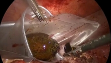
 急性胆囊炎——紧急救治策略
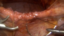
 采用腹腔镜经筋膜腹外缝合术修复因 Morgagn
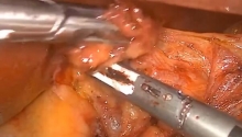
 腹腔镜全胃切除术治疗胃癌中采用线性吻合器
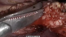
 腹腔镜下Whipple手术治疗胰头肿瘤

 楼主
楼主
 显身卡
显身卡