马上注册,结交更多好友,享用更多功能,让你轻松玩转社区。
您需要 登录 才可以下载或查看,没有账号?注册
×
本帖最后由 小针刀 于 2016-9-7 12:35 编辑
Cowan等人2016年美国移植杂志
Cowan_et_al-2016-American_Journal_of_Transplantation
Incidental Finding in a Renal Transplant Recipient Allograft
肾移植受者的偶发性发现
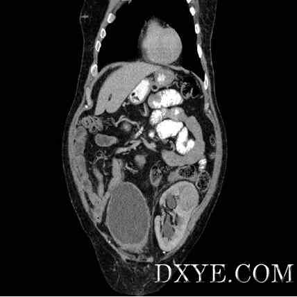
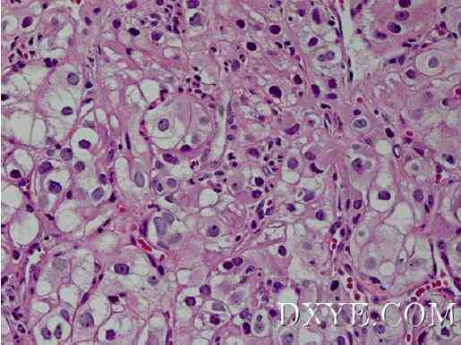
A 67-year-old Caucasian male was referred to our center for the management of transplant hydronephrosis identified on surveillance ultrasound. He had undergone a living related renal transplant 13 years prior to presentation. His medical history was notable for end-stage renal disease due to type 1 diabetes mellitus, and he was maintained on hemodialysis 3 years prior to uneventful transplantation. His serum creatinine at the time of presentation was 1.3 mg/dL (estimated glomerular filtration rate [eGFR] 57 mL/min). A computed tomography (CT) scan with contrast was obtained, and a coronal image is provided (Figure 1). A single renal artery, single renal vein and mild calyceal dilation were identified in addition to an unexpected finding. No prior imaging was available for comparison. A biopsy of this finding was performed, and a representative histologic example is shown (Figure 2). Based on this pathology, the patient was counseled on his treatment options and subsequently taken to the operating room for surgical intervention. His postoperative course was notable for pneumonia, and his serum creatinine 8 months postoperatively was 1.6 mg/dL (eGFR 46 mL/min).
一位67岁的白人男性被称为移植肾积水的超声发现在监控管理中心。介绍他13年前经历了一个与活体有关的肾移植手术。他的病史因终末期肾病而出现,由于1型糖尿病,和他保持着平静移植前3年在血液透析 。他在介绍时的血清肌酐为1.3毫克/升(估计肾小球滤过率[表皮生长因子受体] 57毫升/分钟)。获得了一个对比度的计算机断层扫描(CT),并提供了一个冠状图像(图1)。一个单一的肾动脉、肾静脉和轻度肾盏扩张进行鉴定,除了一个意想不到的发现。没有现有的成像可用于比较。这一发现进行了活检,并显示一个有代表性的组织学例(图2)。基于这一病理,病人接受针对他的治疗方案,随后送往手术室手术干预。他的术后当然是肺炎值得注意的,术后8个月,其血清肌酐水平为1.6毫克/升(表皮生长因子受体46毫升/分钟)。
Figure 1: Coronal computed tomography (CT) image with contrast.

Figure 1: Coronal computed tomography (CT) image with contrast.
图1:冠状计算机体层摄影术(CT)图像与对比。
Figure 2: Representative figure of biopsy histology.

Figure 2: Representative figure of biopsy histology.
图2:活检组织学的代表。
原文:
 Cowan_et_al-2016-American_Journal_of_Transplantation.pdf
(263.37 KB, 下载次数: 0, 售价: 99 香叶)
| 

